Review
. 2016 Feb 25:5:180.
doi: 10.1186/s40064-016-1874-5. eCollection 2016.

Multicentric reticulohistiocytosis (MRH): case report with review of literature between 1991 and 2014 with in depth analysis of various treatment regimens and outcomes

Affiliations
Review

Multicentric reticulohistiocytosis (MRH): case report with review of literature between 1991 and 2014 with in depth analysis of various treatment regimens and outcomes

Saad Tariq et al. Springerplus. .

Abstract

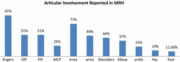
Multicentric reticulohistiocytosis is a rare disease affecting skin and joints primarily and rarely other organs. We present a case report of this disease and an extensive review of the literature. We reviewed the data between 1991 and 2014 and extracted 52 individual cases. Only articles in English were chosen after checking for relevance. The articles were studies and data was extracted into excel spread sheets and later used to compute such variables like frequency, mean and percentage of distribution of various clinical manifestations. The treatments used in these articles were critically analyzed and graded for their relative efficacy for skin and joint manifestations. The grades were 0 = worse, 1 = no benefit/condition remained same, 2 = improvement without resolution, and 3 = resolution. This article also reports the demographic, clinical, laboratory and pathological data from the reviewed articles. Authors attempted to discuss the findings of this review in depth to help manage this condition and proposed a treatment algorithm to help clinicians approach this rare and challenging disease.

Keywords: Arthritis; Autoimmune disease; Immunosuppressive medications; Skin disease.


Figures

Fig. 1
Fig. 1
The patient’s right dorsal hand with visible swelling of all the distal, proximal interphalangeal and metacarpophalangeal joints
Fig. 2
Fig. 2
The right dorsal hand with multiple red-brown macules and papules
Fig. 3
Fig. 3
Skin biopsy from a red-brown papule on the right dorsal hand (Fig. 2) demonstrated numerous multinucleated histiocytes (arrows) infiltrating between the collagen bundles in the superficial dermis. There is a Grenz zone separating the epidermis from the dermal tumor. Additionally, there are an increased number of blood vessels amongst the histiocytes, as well as scattered lymphocytes
Fig. 4
Fig. 4
The multinucleated histiocytes (arrows) are large with an eosinophilic and finely granular “ground-glass” cytoplasm. The nuclei are haphazardly arranged, but tend to favor the center of the cells. A CD163 stain was diffusely positive and the cells were focally PAS-positive diastase-resistant
Fig. 5
Fig. 5
Radiographs of the hands showed erosions in the left third DIP and right second DIP, mild joint space narrowing with mild diffuse osteopenia, and erosions of the scaphoid bone with surrounding soft tissue swelling
Fig. 6
Fig. 6
Radiograph of the left shoulder revealing marked glenohumeral joint space narrowing, small humeral head osteophytes, subchondral sclerosis, and subchondral cysts
Fig. 7
Fig. 7
MRI of the shoulder showing moderate glenohumeral joint effusion with synovitis and large well-demarcated erosions on the humeral head
Fig. 8
Fig. 8
Color Doppler imaging demonstrating a minimal effusion but marked synovial proliferation with moderate Doppler flow
Fig. 9
Fig. 9
Large anechoic effusion and echogenic synovial proliferation with hypervascularity
Fig. 10
Fig. 10
Gender distribution of the cases. Majority of the reported cases were female (71 %) while the rest were male. In few of the cases gender was not clearly reported
Fig. 11
Fig. 11
The most common skin manifestation of MRH was papulonodular rash followed by periungual telengiectasis. Classic coral bead appearance was seen in 28 % cases
Fig. 12
Fig. 12
Hand involvement was the most common region with almost equal involvement of distal and proximal interphalangeal joints. Knee was the second most commonly involved joint with MRH
Fig. 13
Fig. 13
The algorithm summarizes the proposed treatment algorithm. For mild cases NSAIDs can be started but the disease is aggressive in most cases requiring steroids in varying doses and DMARDs. The Methotrexate can be used once the diagnosis is secured with biopsy. Given the erosive nature of the disease with possibility of joint mutilation and good response to biologic agents seen in multiple case reports especially the anti TNF agents, step up therapy should be considered in patients with suboptimal response to DMARDs

References

    1. Adamopoulos IE, Wordsworth PB, Edwards JR, Ferguson DJ, Athanasou NA. Osteoclast differentiation and bone resorption in multicentric reticulohistiocytosis. Hum Pathol. 2006;37(9):1176–1185. doi: 10.1016/j.humpath.2006.04.007. - DOI - PubMed
    1. Aouba A, Leclerc-Mercier S, Fraitag S, Martin-Silva N, Bienvenu B, Georgin-Lavialle S. Assessment and effective targeting of Interleukin-1 in multicentric reticulohistyocytosis. Joint Bone Spine. 2015;82(4):280–283. doi: 10.1016/j.jbspin.2015.02.003. - DOI - PubMed
    1. Barrow MV, Holubar K. Multicentric reticulohistyocitosis. A review of 33 patients. Medicine (Baltimore) 1969;48:287–305. doi: 10.1097/00005792-196907000-00002. - DOI - PubMed
    1. Bennàssar A, Mas A, Guilabert A, Julià M, Mascaró-Galy J, Herrero C. Multicentric reticulohistiocytosis with elevated cytokine serum levels. J Dermatol. 2011;38(9):905–910. - PubMed
    1. Blanco JJ, Hernández FJ, Cerezo JG, García IS, Rubio FC, Rodríguez JJ. Multicentric reticulohistiocytosis. The long course of a rare disease. Scand J Rheumatol. 2002;31(2):107–109. doi: 10.1080/03009740252937658. - DOI - PubMed

LinkOut - more resources