. 1989 Apr 7;57(1):177-87.
doi: 10.1016/0092-8674(89)90183-9.

Genetic control of cell division patterns in the Drosophila embryo

Affiliations

Genetic control of cell division patterns in the Drosophila embryo

B A Edgar et al. Cell. .

Abstract

In Drosophila embryogenesis, mitotic control undergoes a significant transition during the 14th interphase. Mitoses before interphase 14 run on maternal products, and occur in metasynchronous waves. Mitoses after interphase 14 require zygotic transcription, and occur asyncronously in an intricate, highly ordered spatio-temporal pattern. Mutations at the string (stg) locus cause cell-cycle arrest during this transition, in G2 of interphase 14, yet do not arrest other aspects of development. This phenotype suggests that stg is required specifically for initiating mitosis. We describe the cloning of stg, and show that its predicted amino acid sequence is homologous to that of cdc25, a regular of mitotic initiation in the yeast S. pombe. In addition, we show that zygotic expression of stg mRNA occurs in a dynamic series of spatial patterns which anticipate the patterns of the zygotically driven cell divisions. Therefore we suggest that regulated expression of stg mRNA controls the timing and location of these embryonic cell divisions.


Figures

Figure 1
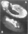
Figure 1. Wild-Type (A) and stg (B) Embryos at about 6 Hr AED, Stained with Anti-Tubulin Antibodies
Anterior is to the left and dorsal is up. Tubulin outlines the cells during interphase, and forms spindles (which appear as bright dots) during mitosis. Note that the stg embryo has many fewer cells than the wild-type embryo. The wild-type embryo is undergoing mitosis 15, whereas the stg embryo is arrested in interphase 14. Other morphological features, such as the extended germ band, gnathal lobes, and amnioserosa, are similar in both embryos.
Figure 2
Figure 2. DNA Synthesis in (A) Wild-Type and (B) stg7B69 Embryos
Anterior is to the left and dorsal up. We injected these embryos with 50 mM BrdU (pH 9) during G2 of cycle 14, allowed them to develop for 3 hr, and then fixed them. BrdU-labeled DNA was detected by indirect immunofluorescence (Schubiger and Palka, 1987). All nuclei in the wild-type embryo are labeled, whereas only the polyploid nuclei of the amnioserosa are labeled in the stg7B69 embryo. In the wild-type embryo (A), the amnioserosa nuclei are polyploid, but are not heavily labeled. This is probably due to depletion of the injected BrdU prior to polyploidization.
Figure 3
Figure 3. Cloning of the stg Locus
The figure shows a restriction map of the 33 kb genomic region that contains the stg locus, with EcoRI (r) and SaII (s) sites indicated. The insertion site of the P element (pUChsneo; Steller and Pirotta, 1985) in stg mutants I(3)-neo61 and I(3)neo62 is indicated above the map. We isolated 4.5 kb of DNA flanking these insertions by plasmid rescue (p62s) and used this to isolate genomic clones (λ1, λ3b) from a lambda library. The entire region was fragmented into nine contiguous pieces (a–i), and the isolated fragments were used to probe developmental Northern blots, examples of which are shown below the map. Probes a–d detected no transcripts, probes e and f detected transcripts of 2.8 and 3.0 kb, probe g detected a 2.9 kb transcript, and probes h and i detected transcripts of 2.9 and 2.7 kb. The inferred transcribed regions are indicated by thin lines below the probe fragments, and the direction of transcription of the presumed stg transcripts is indicated with an arrow. Numbers below the blots indicate hr AED at 22°C. One-hour embryos (1) are rapidly cleaving, 3 hr embryos (3) are in interphase 14, 5 hr embryos (5) are undergoing mitoses 14 and 15, and 12 hr embryos (12) have only a few mitoses, mainly in the nervous system. Above the restriction map we show a Southern blot of genomic DNA from the various P element mutants and revertants, cut with EcoRI and hybridized with probe d, a 2.2 kb genomic EcoRI fragment. Numbers above the gel panel indicate genotypes: (1) I(3)neo31/TM3 (a P element insertion at a different site, this is a control); (2) I(3)neo62/TM3; (3) revertant neo62R2/neo62R2; (4) revertant neo62R5/neo62R5; (5) I(3)3A1/TM3; (6) I(3)1D3/TM3. Sizes of the bands detected are 3.6, 2.2, and 2.1 kb, top to bottom. The wild-type band is 2.2 kb (lane 1). In each of the P element insertion mutants, this band is altered in size (lanes 2, 5, 6), and in the P element revertants it is restored to approximately the wild-type size (lanes 3 and 4).
Figure 4
Figure 4. Nucleotide and Predicted Amino Acid Sequence of a 2308 Base stg cDNA That Is Homologous to Genomic Fragments e and f (Figure 3)
The translation initiation site was chosen to maximize the length of the open reading frame. Both this site and one at position 31 match the consensus sequence for translation initiation sites in Drosophila (Cavener, 1987).
Figure 5
Figure 5. The Predicted Amino Acid Sequence of stg Is Homologous to cdc25 of S. pombe (25) and MIH1 of S. cerevisiae (mih)
The C-terminal regions of the three sequences are aligned, and identical amino acids are boxed. The three sequences are approximately 35% identical in a 187 amino acid stretch included in the figure. We found no significant homologies in the N-terminal regions of these proteins.
Figure 6
Figure 6. A Temporal Sequence of stg mRNA Expression Patterns during Cycles 14 and Early 15, Spanning about 2 hr of Development (25°)
The right column shows stg mRNA patterns, visualized by in situ hybridization of a stg cDNA probe to whole embryos. The left column shows the same embryos stained for DNA with Hoechst 33258. Embryos A-D were hybridized with 35S probe, which detects expression through several layers of tissue. Embryos E-H were hybridized with 3H probe, which detects only surface expression. The anterior of each embryo is to the left. We have labeled the regions of stg RNA expression with the numbers of the mitotic domains (MDs) mapped by Foe (1989). stg expression corresponds perfectly with these domains, but begins 25–35 min before the onset of mitosis in a given domain. A: ventral view, showing the first zygotic stg expression in MD10 (10), and what is probably residual maternal RNA in the pole cells (P). B: dorsal view, just at the onset of gastrulation, about 25 min before the first mitoses. stg expression can be seen in MDs 1–10. C: dorsal view showing the beginning of expression in five spots within MD11, about 15 min before the first mitoses. D: dorsolateral view, showing expanded expression in MD11. The first mitoses, in MDs 1–8, are in progress in this embryo. E: ventrolateral view of an embryo the same age as D. stg expression can be seen in MDs 1, 2, 5, 8, 9, 11, 14, and 15. Mitoses are occurring in domains 1, 2, 5, 8, and 9, but have not yet begun in MDs 11, 14, or 15. The embryo just above this (not labeled) is slightly older and shows segmentally repeated spots of expression in MDs 16 and 17. F: ventrolateral view showing stg expression and mitoses in MD N. Note that expression has diminished in MDs 1–15, which have already divided. Also note the lack of expression in the amnioserosa (A), a nondividing polyploid tissue. G: lateral view, showing cycle 14 expression in MDs N and M, which are dividing. Cycle 15 expression is evident in a number of spots in the head, and in a segmental pattern in the dorsolateral epidermis and mesectoderm.
Figure 7
Figure 7. An Example of the Correlation of stg RNA Expression with the Mitotic Pattern
A: A whole, late cycle 14 embryo hybridized with 3H-labeled stg cDNA probe. The photograph is a double exposure: silver grains show up as white dots and nuclei stained with Hoechst 33258 are gray. Arrows indicate pairs of adjacent cells containing very different levels of stg RNA. B: A tracing of the embryo in A, showing the correlation of stg expression domains (shaded) with the mitotic domains (numbers). These numbers were assigned by Foe (1989) to each mitotic domain, and indicate the relative timing of mitotic initiation in different domains. Note that stg RNA levels are highest in the earliest dividing domains (1–8), and lower in later dividing domains (9–16). This embryo has initiated mitosis in domains 1–6. C: A slightly older embryo stained with anti-tubulin antibodies to show mitotic spindles. In this embryo divisions have already occured in mitotic domains 1–8, and are in progress in domains 9, 11, 14, and 15 (numbers). Mitotic domains 10, 12, and 13 are not visible from this view. Small arrows indicate adjacent cells with different mitotic states. All embryos are mounted with anterior to the left and dorsal uppermost.

References

    1. Arion D, Meijer L, Brizuela L, Beach D. cdc2 is a component of the M phase–specific histone H1 kinase: evidence for identity with MPF. Cell. 1988;55:371–378. - PubMed
    1. Berger C. In situ hybridization of immunoglobulin-specific RNA in single cells of the B lymphocyte lineage with radiolabeled DNA probes. EMBO J. 1986;5:85–93. - PMC - PubMed
    1. Blow JJ, Laskey RA. A role for the nuclear envelope in controlling DNA replication within the cell cycle. Nature. 1988;332:546–548. - PubMed
    1. Blumenthal AB, Kriegstein HJ, Hogness DS. The units of DNA replication in Drosophila melanogaster chromosomes. Cold Spring Harbor Symp Quant Biol. 1974;38:205–223. - PubMed
    1. Cavener DR. Comparison of the consensus sequence flanking translational start sites in Drosophila and vertebrates. Nucl Acids Res. 1987;15:1353–1361. - PMC - PubMed

Publication types

MeSH terms

Associated data