

PROSPECTIVE ASSESSMENT OF THE CLINICAL, RADIOGRAPHIC AND FUNCTIONAL EVOLUTION OF TREATMENT FOR UNSTABLE TROCHANTERIC FRACTURES OF THE FEMUR USING A CEPHALOMEDULLARY NAIL
- PMID: 27027025
- PMCID: PMC4799315
- DOI: 10.1016/S2255-4971(15)30249-4
PROSPECTIVE ASSESSMENT OF THE CLINICAL, RADIOGRAPHIC AND FUNCTIONAL EVOLUTION OF TREATMENT FOR UNSTABLE TROCHANTERIC FRACTURES OF THE FEMUR USING A CEPHALOMEDULLARY NAIL
Abstract
To assess the clinical, radiological and functional evolution of osteosynthesis using a cephalomedullary nail, in unstable trochanteric fractures of the femur, over a one-year postoperative follow-up.
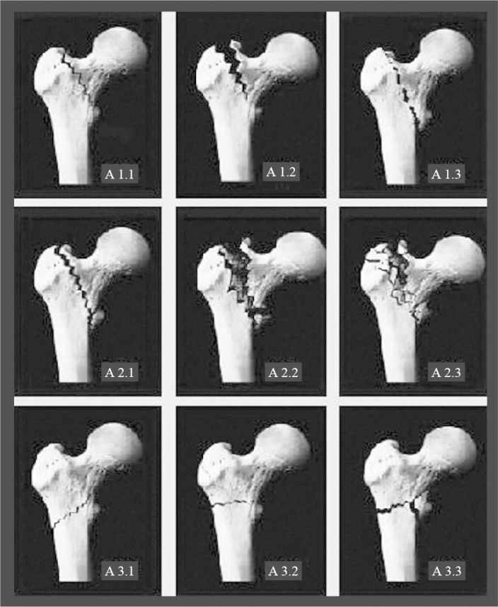
Methods: Fourteen men and 23 women of mean age 77.7 years were evaluated. Twenty-seven of them had fractures classified as AO/ASIF 31A2 and ten as 31A3. The patients were evaluated clinically, radiologically and functionally one week, two weeks, one month, two months, six months and one year after the operation.
Results: The clinical complications comprised five cases of death, one case of calcaneal ulcer, one case of acute arterial obstruction and two cases of deep vein thrombosis. The radiographic evaluation showed that the mean cervicodiaphyseal angle in the immediate postoperative period was 132.5°. The mean tip-apex index was 22.8 mm. After one year, the mean cervicodiaphyseal angle was 131.7°. Fracture consolidation was seen in all the patients six months after the operation, except in one case that presented cut-out. There were no cases of fracture below the implant. The functional evaluation using the Harris score after one year showed a mean of 69.3 points. The evaluation of walking progress showed that after one year, 40.6% of the patients had the same ability to walk that they had before the fracture. The visual analogue pain scale showed that a significant decrease in pain complaints occurred, going from 5.19 in the first week to 2.25 after 1 year.
Conclusion: Osteosynthesis using a cephalomedullary nail resulted in low rates of clinical and mechanical complications and adequate functional outcomes.
Keywords: Bone Nails; Femoral Fractures; Fracture Fixation; Fracture Healing; Hip Fractures; Internal/methods; Postoperative Complications.
Figures


References
-
- Parker MJ, Handoll HH. Intramedullary nails for extracapsular hip fractures in adults. Cochrane Database Syst Rev. 2009;(3):CD004961. - PubMed
-
- Kaplan K, Miyamoto R, Levine BR, Egol KA, Zuckerman JD. Surgical management of hip fractures: an evidence-based review of the literature. II. Intertrochanteric fractures. J Am Acad Orthop Surg. 2008;16(11):665–673. - PubMed
-
- Haidukewych GJ. Intertrochanteric fractures: ten tips to improve results. J Bone Joint Surg Am. 2009;91(3):712–719. - PubMed
-
- Müller ME. Classification and international AO-Documentation of femur fractures. Unfallheilkunde. 1980;83(5):251–259. - PubMed
LinkOut - more resources
Full Text Sources
Miscellaneous