

PHYSICOCHEMICAL CHARACTERIZATION OF LYOPHILIZED BOVINE BONE GRAFTS
- PMID: 27027036
- PMCID: PMC4799317
- DOI: 10.1016/S2255-4971(15)30260-3
PHYSICOCHEMICAL CHARACTERIZATION OF LYOPHILIZED BOVINE BONE GRAFTS
Abstract
To evaluate the physicochemical characteristics of lyophilized bovine grafts manufactured on a semi-industrial scale (Orthogen; Baumer S/A*) in accordance with a protocol previously developed by the authors.
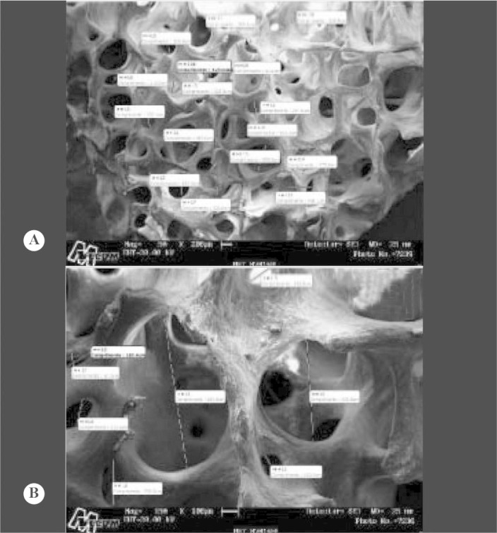
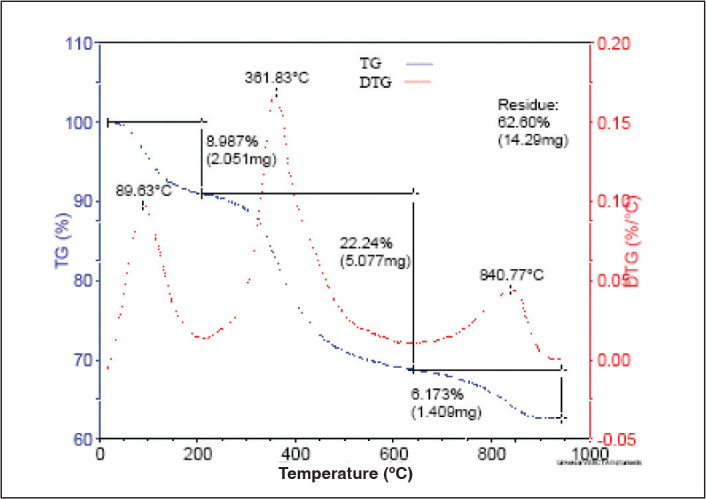
Methods: The lyophilized bovine bone grafts were characterized by means of scanning electron microscopy (SEM), energy dispersive spectroscopy (EDS), X-ray diffractometry (XRD), thermogravimetric (TG) analysis, differential exploratory scanning calorimetry (DSC) and Fourier-transform infrared (FT-IR) spectroscopy.
Results: Ca was the main component (60%) found in the samples, followed by P (28%) and O (5%). The mean (sd) pore size was 316 μm (146.7), ranging from 91.2 to 497.8 μm, and 333.5 μm (304.8), ranging from 87.2 to 963.9 μm, at 50x and 150x magnification, respectively. The hydroxyapatite peaks were at 26°C and 32°C, and mass losses were observed between 250°C and 640°C, corresponding to organic material and water. Two temperature transitions (45.67°C and 91.89°C) showed denaturation of type 1 collagen and dehydration of hydroxyapatite.
Conclusion: The physicochemical assessment of lyophilized bovine bone grafts in accordance with the protocol developed at semi-industrial scale confirmed that this product presents excellent biocompatibility, with characteristics similar to natural bone.
Keywords: Biocompatible Materials; Bone Transplantation; General Surgery.
Figures


References
-
- Finkemeier CG. Bone-grafting and bone-graft substitutes. J Bone Joint Surg Am. 2002;84(3):454–464. - PubMed
-
- Laurencin CT, Khan Y. Bone grafts and bone graft substitutes: a brief history. In: Laurencin CT, editor. Bone graft substitutes. ASTM International; Bridfeport, NJ: 2003.
-
- Seiler JG 3rd, Johnson J, Hand G, Microsurgery clinic. Iliac crest autogenous bone grafting: donor site complications. J South Orthop Assoc. [periódico online]. Disponível em: http://www.medscape.com/viewarticle/410431. Acesso em: 11 setembro, 2002 - PubMed
-
- Lind M, Krarup N, Mikkelsen S, Hørlyck E. Exchange impaction allografting for femoral revision hip arthroplasty: results in 87 cases after 3.6 years' follow-up. J Arthroplasty. 2002;17(2):158–164. - PubMed
-
- Palmer SH, Gibbons CL, Athanasou NA. The pathology of bone allograft. J Bone Joint Surg Br. 1999;81(2):333–335. - PubMed
LinkOut - more resources
Full Text Sources
Miscellaneous