

UPDATING ON DIAGNOSIS AND TREATMENT OF CHONDRAL LESION OF THE KNEE
- PMID: 27027078
- PMCID: PMC4799341
- DOI: 10.1016/S2255-4971(15)30339-6
UPDATING ON DIAGNOSIS AND TREATMENT OF CHONDRAL LESION OF THE KNEE
Abstract
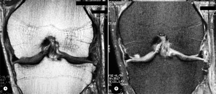
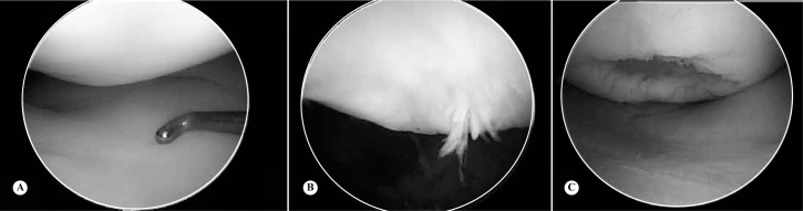
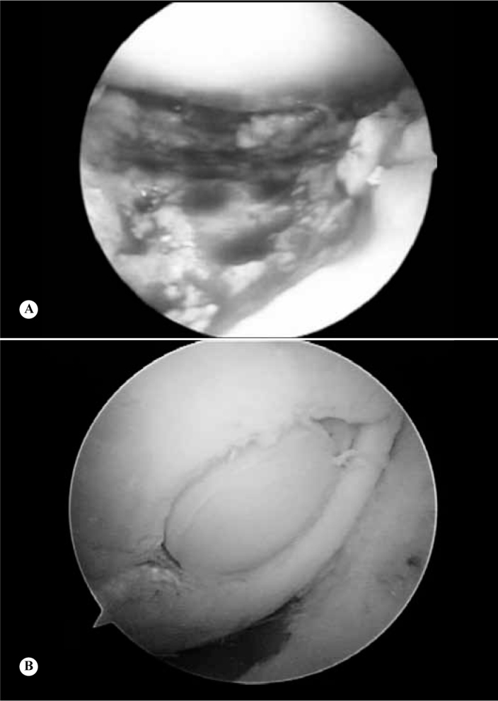
The treatment of chondral knee injuries remains a challenge for the orthopedic surgeon, mainly owing to the characteristics of the cartilage tissue, which promote low potential for regeneration. Chondral lesions can be caused by metabolic stimulation, or by genetic, vascular and traumatic events, and are classified according to the size and thickness of the affected cartilage. Clinical diagnosis can be difficult, especially due to insidious symptoms. Additional tests, as Magnetic Resonance Imaging (MRI), may be needed. The treatment of these lesions usually starts with non-operative management. Surgery should be reserved for patients with detached chondral fragments, blocked range of motion, or the failure of non-operative treatment. The surgical techniques used for the treatment of partial thickness defects are Debridement and Ablation. These techniques aim to improve symptoms, since they do not restore normal structure and function of the cartilage. For full-thickness defects (osteochondral lesion), available treatments are Abrasion, Drilling, Microfracture, Osteochondral Autologous and Allogeneic Transplantation, and biological techniques such as the use of Autologous Chondrocyte Transplantation, Minced Cartilage and stem cells.
Keywords: Arthroscopy; Articular Cartilage; Cartilage Diseases; Knee.
Figures
References
-
- Blackburn TA, Craig E. Knee anatomy: a brief review. Phys Ther. 1980;60(12):1556–1560. - PubMed
-
- Ferretti M, Viola DCM, Garcia RJ. Tratamento dos defeitos osteocondrais. Einstein. 2009;7(2 Pt 1):245–247.
-
- Aigner T, Stöve J. Collagens–major component of the physiological cartilage matrix, major target of cartilage degeneration, major tool in cartilage repair. Adv Drug Deliv Rev. 2003;55(12):1569–1593. - PubMed
-
- Rosowski M, Falb M, Tschirschmann M, Lauster R. Initiation of mesenchymal condensation in alginate hollow spheres–a useful model for understanding cartilage repair? Artif Organs. 2006;30(10):775–784. - PubMed
-
- Richmond JC. Surgery for osteoarthritis of the knee. Rheum Dis Clin North Am. 2008;34(3):815–825. - PubMed
LinkOut - more resources
Full Text Sources
Medical