Review
. 2016 Sep 1;25(7):373-88.
doi: 10.1089/ars.2015.6493. Epub 2016 Mar 30.

Endothelial Mechanosignaling: Does One Sensor Fit All?

Affiliations
Review

Endothelial Mechanosignaling: Does One Sensor Fit All?

Chris Givens et al. Antioxid Redox Signal. .

Abstract

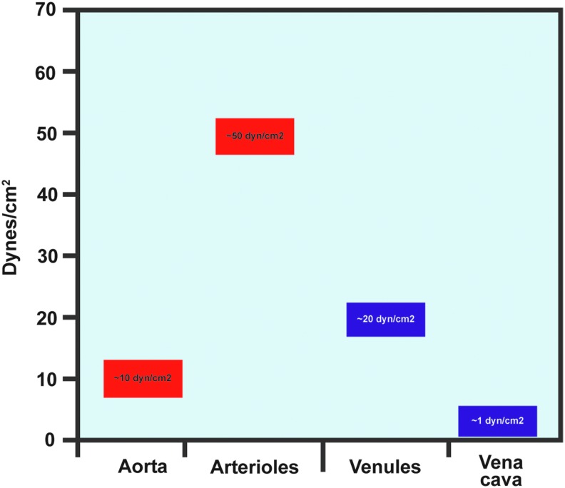
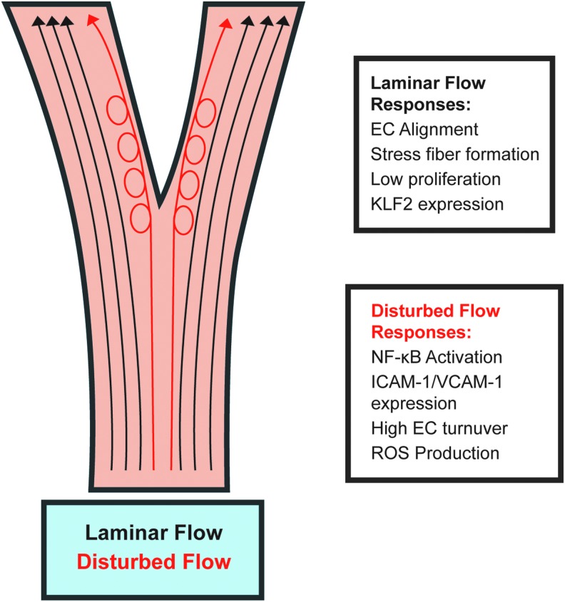
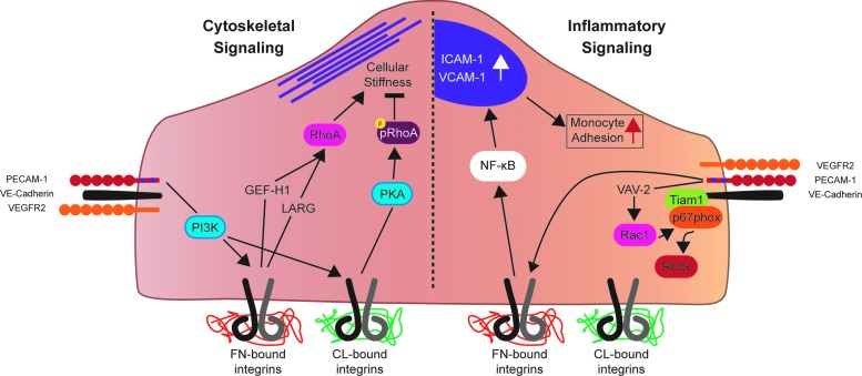
Significance: Forces are important in the cardiovascular system, acting as regulators of vascular physiology and pathology. Residing at the blood vessel interface, cells (endothelial cell, EC) are constantly exposed to vascular forces, including shear stress. Shear stress is the frictional force exerted by blood flow, and its patterns differ based on vessel geometry and type. These patterns range from uniform laminar flow to nonuniform disturbed flow. Although ECs sense and differentially respond to flow patterns unique to their microenvironment, the mechanisms underlying endothelial mechanosensing remain incompletely understood.

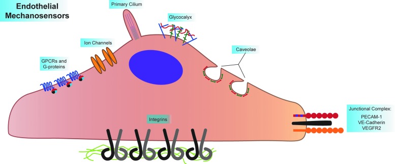
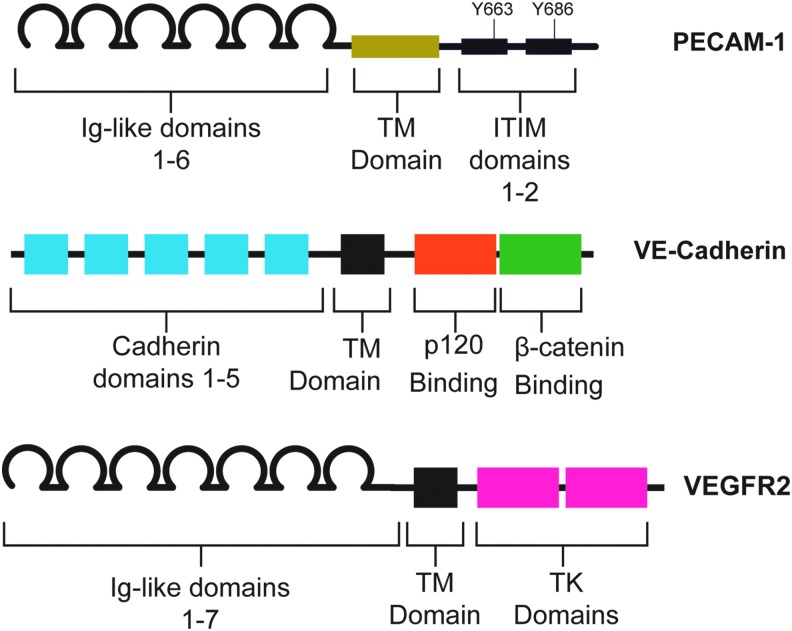
Recent advances: A large body of work suggests that ECs possess many mechanosensors that decorate their apical, junctional, and basal surfaces. These potential mechanosensors sense blood flow, translating physical force into biochemical signaling events.

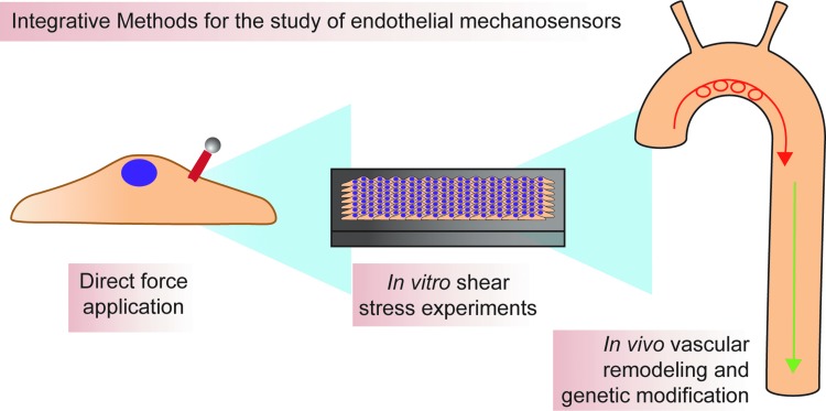
Critical issues: Understanding the mechanisms by which proposed mechanosensors sense and respond to shear stress requires an integrative approach. It is also critical to understand the role of these mechanosensors not only during embryonic development but also in the different vascular beds in the adult. Possible cross talk and integration of mechanosensing via the various mechanosensors remain a challenge.

Future directions: Determination of the hierarchy of endothelial mechanosensors is critical for future work, as is determination of the extent to which mechanosensors work together to achieve force-dependent signaling. The role and primary sensors of shear stress during development also remain an open question. Finally, integrative approaches must be used to determine absolute mechanosensory function of potential mechanosensors. Antioxid. Redox Signal. 25, 373-388.


Figures

<b>FIG. 1.</b>
FIG. 1.
Shear stress levels are variable throughout the vasculature. Arterial shear stress levels are higher than venous levels, and larger vessels have lower shear than smaller vessels. To see this illustration in color, the reader is referred to the web version of this article at www.liebertpub.com/ars
<b>FIG. 2.</b>
FIG. 2.
Two main types of flow exist in the vasculature: laminar and disturbed flow. Laminar flow occurs where vessel gemoetry is straight and uniform, whereas disturbed flow occurs where vessels bifurcate or curve highly. The different types of flow elicit different endothelial responses. Responses to laminar flow, termed “atheroprotective,” include EC alignment in the direction of flow, stress fiber formation, and KLF2 expression, which lead to anti-inflammatory gene expression. Disturbed flow responses, termed “atheroprone,” are more inflammatory and include NF-κB activation and associated transcription. In addition, ECs in areas of disturbed flow are more proliferative and produce more ROS than ECs in areas of laminar flow. EC, endothelial cell; ROS, reactive oxygen species. To see this illustration in color, the reader is referred to the web version of this article at www.liebertpub.com/ars
<b>FIG. 3.</b>
FIG. 3.
Endothelial cells express many mechanosensors. These sensors can be divided into luminal, junctional, and basal mechanosensors. Luminal mechanosensors include G-protein-coupled receptors (GPCRs, including the sphingosine 1-phosphate receptor 1, S1P1, and the bradykinin B2 receptor) and heterotrimeric G-proteins (namely, Gαq/11), ion channels (including TRPV4, TRPP2, TRPC1, Piezo1, and Piezo2), microtubule-based primary cilia (which associated with the ion channels PKD1 and PKD1), the glycocalyx (where Syndecan-1 and −4, as well as heparin sulfate glycosaminoglycans are involved in shear signaling), and protein-coated membrane pits called caveolae (given structure by Caveolin 1–3 and Cavin 1–3). The known junctional mechanosensors, PECAM-1, VE-Cadherin, and VEGFR2, form a mechanosensory complex that elicits many signaling pathways as a response to shear. The basal mechanosensors consist of the integrins, which sense ECM type and substrate stiffness, all while integrating signaling pathways originating from other mechanosensors. ECM, extracellular matrix; GPCR, G-protein-coupled receptors; PECAM-1, platelet endothelial cell adhesion molecule-1. To see this illustration in color, the reader is referred to the web version of this article at www.liebertpub.com/ars
<b>FIG. 4.</b>
FIG. 4.
Domain structure of the junctional mechanosensors. The extracellular domain of PECAM-1 comprises six Ig-like repeats, which participate in homophilic binding with other PECAM-1 molecules on neighboring ECs. These Ig-like repeats are linked by a TM domain to the intracellular domain. The intracellular domain contains two ITIM domains, which contain critical tyrosines that are rapidly phosphorylated at the onset of shear. VE-Cadherin, a classical cadherin, has an extracellular domain that comprised five cadherin domains, which mediate adhesion, and features p120 and β-catenin binding sites on its intracellular domain. These intracellular domains interact with p120 and β-catenin, mediating VE-Cadherin binding to the cytoskeleton. VEGFR2, a receptor tyrosine kinase has seven Ig-like repeats in its extracellular domain. The intracellular domain contains two TK domains that interact with downstream effectors. Critically, tyrosines 801 and 1175 bind PI3K upon stimulation of the receptor. Mutation of these tyrosines blunts the cellular response to shear. Ig, immunoglobulin; ITIM, immunoreceptor tyrosine-based inhibitory motif; TK, tyrosine kinase; TM, transmembrane. To see this illustration in color, the reader is referred to the web version of this article at www.liebertpub.com/ars
<b>FIG. 5.</b>
FIG. 5.
The junctional mechanosensory complex regulates cytoskeletal stiffening and inflammatory signaling in response to force. Force on PECAM-1 activates PI3K, which is required for downstream activation of integrins. ECM-dependent integrin activation occurs, with FN bound integrins positively regulating cellular stiffness through GEF-H1/LARG-RhoA signaling. CL bound integrins have the opposite affect; cellular stiffness is negatively regulated downstream of CL-bound integrins through a PKA-phospho-RhoA pathway. In addition, Rac1 is activated downstream of force on PECAM-1, which leads to increased inflamatory ROS production. In addition, shear stress activates FN-bound integrins, leading to NF-κB activation, ICAM-1 and VCAM-1 expression, and an increase in moncyte adhesion to “activated” ECs. CL, collagen; FN, fibronectin; PI3K, phosphoinositide 3-kinase; PKA, protein kinase A. To see this illustration in color, the reader is referred to the web version of this article at www.liebertpub.com/ars
<b>FIG. 6.</b>
FIG. 6.
Integration of approaches is necessary for determining the precise function of various mechanosensors. Methods available for the study of mechanosensing range from reductionist biophysical experiments to in vivo hemodynamic modification. Magnetic beads are used to apply force directly to putative mechanosensors and can be useful for directly determing mechanosensitivity of a protein. In vitro shear stress experiments are useful for determining mechanoresponsiveness of proteins in the context of an endothelial monolayer, while in vivo experiments determine whether information gained using the aforementioned reductionist approaches is physiologically relevant. To see this illustration in color, the reader is referred to the web version of this article at www.liebertpub.com/ars

References

    1. AbouAlaiwi WA, Takahashi M, Mell BR, Jones TJ, Ratnam S, Kolb RJ, and Nauli SM. Ciliary polycystin-2 is a mechanosensitive calcium channel involved in nitric oxide signaling cascades. Circ Res 104: 860–869, 2009 - PMC - PubMed
    1. Alexander CM, Reichsman F, Hinkes MT, Lincecum J, Becker KA, Cumberledge S, and Bernfield M. Syndecan-1 is required for Wnt-1-induced mammary tumorigenesis in mice. Nat Genet 25: 329–332, 2000 - PubMed
    1. Ando J, Ohtsuka A, Korenaga R, Kawamura T, and Kamiya A. Wall shear stress rather than shear rate regulates cytoplasmic Ca++ responses to flow in vascular endothelial cells. Biochem Biophys Res Commun 190: 716–723, 1993 - PubMed
    1. Ando J. and Yamamoto K. Effects of shear stress and stretch on endothelial function. Antioxid Redox Signal 15: 1389–1403, 2011 - PubMed
    1. Baeyens N, Mulligan-Kehoe MJ, Corti F, Simon DD, Ross TD, Rhodes JM, Wang TZ, Mejean CO, Simons M, Humphrey J, and Schwartz MA. Syndecan 4 is required for endothelial alignment in flow and atheroprotective signaling. Proc Natl Acad Sci USA 111: 17308–17313, 2014 - PMC - PubMed

Publication types

LinkOut - more resources