. 2016 Jun 14;7(24):35618-35631.
doi: 10.18632/oncotarget.8343.

Poly(ADP-ribose) polymerase 1 inhibition protects cardiomyocytes from inflammation and apoptosis in diabetic cardiomyopathy

Affiliations

Poly(ADP-ribose) polymerase 1 inhibition protects cardiomyocytes from inflammation and apoptosis in diabetic cardiomyopathy

Wei-Dong Qin et al. Oncotarget. .

Abstract

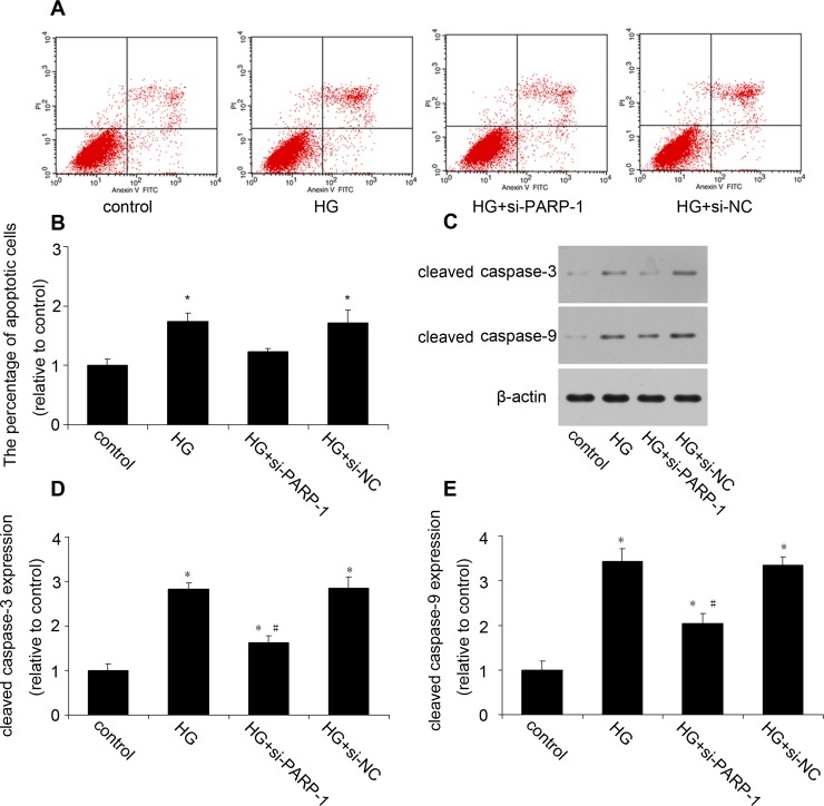
Diabetic cardiomyopathy (DCM) is characterized by structural alterations such as cardiomyocyte hypertrophy, necrosis and focal fibrosis. Poly(ADP-ribose) polymerase 1 (PARP-1) is a nuclear enzyme which can be activated by DNA damage and plays a critical role in various diseases. We hypothesized that PARP-1 may play an important role in DCM and that its inhibition may protect cardiomyocytes from inflammation and apoptosis in DCM. H9c2 cardiomyocytes were treated with normal glucose, mannitol or high glucose (HG). Male C57BL/6 mice or PARP-1-/- mice were treated with streptozotocin (STZ) by intraperitoneal injection for 5 consecutive days to induce diabetes. In vitro, HG stimulation induced oxidative stress and DNA damage and increased PARP-1 expression and activity. Compared with the control, pretreatment with PARP-1 siRNA significantly reduced HG-induced inflammatory response, including tumor necrosis factor-α (TNF-α), interleukin-1β (IL-1β) and IL-6 secretion, and intercellular adhesion molecule-1 (ICAM-1) and inducible nitric oxide synthase (iNOS) expression. PARP-1 inhibition reduced HG-induced cardiomyocyte apoptosis through downregulation of cleaved caspases and activation of IGF-1R/Akt pathway. In vivo, hyperglycemia increased the protein expression of nitrotyrosine and PARP-1 as well as PARP-1 activity. PARP-1 gene deletion significantly improved cardiac dysfunction and reduced inflammatory response and apoptosis. This work demonstrated the critical role of PARP-1 in diabetic heart injury, and suggested that PARP-1 inhibition may be a feasible strategy for the treatment of DCM.

Keywords: Pathology Section; apoptosis; diabetic cardiomyopathy; hyperglycemia; inflammatory response; poly(ADP-ribose) polymerase 1.


Conflict of interest statement

No conflict of interest for all authors.

Figures

Figure 1
Figure 1. HG increased PARP-1 expression and activity and induced DNA damage
After H9c2 cells were stimulated by normal glucose (control, 5.5 mmol/l glucose), mannitol (5.5 mmol/l glucose plus 24.5 mmol/l mannitol) or high glucose (HG, 33mmol/l glucose), PARP-1 expression and activity was assessed, oxidative stress was determined by DHE and DCF staining, and DNA damage was assessed by comet assay. A. HG simulation increased PARP-1 mRNA expression as determined by RT-PCR. B., C. HG simulation increased PARP-1 protein expression as assessed by western blotting analysis. D. HG simulation increased PARP-1 activity as determined by spectrophotometer. E., F., G. HG simulation induced oxidative stress. H., I. HG treatment induced DNA damage. Values are expressed as mean ± S.D. from three independent experiments. *P < 0.05 vs. control. #P < 0.05 vs. 24h. HG: high glucose.
Figure 2
Figure 2. PARP-1 inhibition reduced HG-induced inflammatory response
H9c2 cells were stimulated by normal glucose or HG after PARP-1 was inhibited by siRNA for 24hrs. The secretion of TNF-α, IL-1β and IL-6 was determined by ELISA, the expression of ICAM-1 and iNOS was assessed by RT-PCR and western blotting analysis. A., B., C. PARP-1 inhibition by siRNA reduced HG-induced the cellular secretion of TNF-α, IL-1β and IL-6. D., E. PARP-1 inhibition reduced HG-induced the mRNA expression of ICAM-1 and iNOS. F., G., H. PARP-1 inhibition reduced HG-induced the protein expression of ICAM-1 and iNOS. Values are expressed as mean ± S.D. *P < 0.05 vs. control. #P < 0.05 vs. HG. HG: high glucose; si-PARP-1: PARP-1 siRNA; si-NC: negative control of PARP-1 siRNA.
Figure 3
Figure 3. PARP-1 inhibition reduced HG-induced cell apoptosis
H9c2 cells were stimulated by normal glucose or HG after PARP-1 was inhibited by siRNA for 24hrs. Cell apoptosis was assessed by flow cytometry and cleavaged caspase was detected by western blotting analysis. A., B. PARP-1 inhibition reduced HG-induced cell apoptosis. C., D., E. PARP-1 inhibition reduced HG-upregulated the protein expression of cleaved caspase-3 and caspase-9. Values are expressed as mean ± S.D. *P < 0.05 vs. control. #P < 0.05 vs. HG. HG: high glucose; si-PARP-1: PARP-1 siRNA; si-NC: negative control of PARP-1 siRNA.
Figure 4
Figure 4. PARP-1 inhibition increased HG-reduced the phosphorylation of IGF-1R/Akt
H9c2 cells were stimulated by normal glucose or HG after PARP-1 was inhibited by siRNA for 24hrs. The protein expression of phosphorylated IGF-1R and Akt was detected by western blotting analysis. A., B. PARP-1 inhibition increased HG-reduced the phosphorylation of IGF-1R. C., D. PARP-1 inhibition increased HG-reduced the phosphorylation of Akt. Values are expressed as mean ± S.D. *P < 0.05 vs. control. #P < 0.05 vs. HG. HG: high glucose; si-PARP-1: PARP-1 siRNA; si-NC: negative control of PARP-1 siRNA.
Figure 5
Figure 5. PARP-1 inhibition improved hyperglycemia-reduced cardiac function
Before mice were sacrified, cardiac diameter and function were measured by Echocardiography. Compared with the control, hyperglycemia increased LVPWd and reduced LVEF, FS and E/A in DM mice, while PARP-1 deletion alleviated these effects. LV: Left ventricular; LVEF: left ventricular ejection fraction; FS: fractional shortening; E/A: ratio of early to late mitral inflow velocity; LVEDd: left ventricular end-diastolic dimension. Values are expressed as mean ± S.D. *P < 0.05 vs. control. DM: diabetic cardiomyopathy.
Figure 6
Figure 6. Myocardial pathology feature and PARP-1 expression in mice
A. PARP-1 deletion improved hyperglycemia-induced cardiac remodeling. B., C., D. Hyperglycemia increased the protein expression of nitrotyrosine and PARP-1. E. Hyperglycemia increased PARP-1 activity. Values are expressed as mean ± S.D. *P < 0.05 vs. control. DM: diabetic cardiomyopathy; si-PARP-1: PARP-1 siRNA; si-NC: negative control of PARP-1 siRNA.
Figure 7
Figure 7. PARP-1 deletion reduced hyperglycemia-induced inflammatory response in mice
In mice, the plasma concentration of TNF-α, IL-1β and IL-6 was determined by ELISA, the expression of ICAM-1 and iNOS was assessed by RT-PCR and western blotting analysis. A., B., C. PARP-1 deletion reduced hyperglycemia-upregulated plasma concentration of TNF-α, IL-1β and IL-6. D., E. PARP-1 deletion reduced hyperglycemia-upregulated the mRNA expression of ICAM-1 and iNOS. F., G., H. PARP-1 deletion reduced hyperglycemia-upregulated the protein expression of ICAM-1 and iNOS. Values are expressed as mean ± S.D. *P < 0.05 vs. control. #P < 0.05 vs. DM. DM: diabetic cardiomyopathy.
Figure 8
Figure 8. PARP-1 deletion reduced hyperglycemia-induced cardiomyocyte apoptosis
A., B. PARP-1 deletion reduced hyperglycemia-induced cardiomyocyte apoptosis. C., D., E. PARP-1 deletion reduced hyperglycemia-upregulated the protein expression of cleaved caspase-3 and caspase-9. F., G., H. PARP-1 deletion increased hyperglycemia-reduced the phosphorylation of IGF-1R/Akt. Values are expressed as mean ± S.D. *P < 0.05 vs. control. #P < 0.05 vs. DM. DM: diabetic cardiomyopathy.

References

    1. Pieper AA, Brat DJ, Krug DK, Watkins CC, Gupta A, Blackshaw S, Verma A, Wang ZQ, Snyder SH. Poly(adp-ribose) polymerase-deficient mice are protected from streptozotocin-induced diabetes. Proc Natl Acad Sci U S A. 1999;96:3059–3064. - PMC - PubMed
    1. Ti Y, Xie GL, Wang ZH, Bi XL, Ding WY, Wang J, Jiang GH, Bu PL, Zhang Y, Zhong M, Zhang W. Trb3 gene silencing alleviates diabetic cardiomyopathy in a type 2 diabetic rat model. Diabetes. 2011;60:2963–2974. - PMC - PubMed
    1. Kanda T, Hayashi K, Wakino S, Homma K, Yoshioka K, Hasegawa K, Sugano N, Tatematsu S, Takamatsu I, Mitsuhashi T, Saruta T. Role of rho-kinase and p27 in angiotensin ii-induced vascular injury. Hypertension. 2005;45:724–729. - PubMed
    1. Wan X, Yeung C, Heske C, Mendoza A, Helman LJ. Igf-1r inhibition activates a yes/sfk bypass resistance pathway: Rational basis for co-targeting igf-1r and yes/sfk kinase in rhabdomyosarcoma. Neoplasia. 2015;17:358–366. - PMC - PubMed
    1. Westermann D, Van Linthout S, Dhayat S, Dhayat N, Escher F, Bücker-Gärtner C, Spillmann F, Noutsias M, Riad A, Schultheiss HP, Tschöpe C. Cardioprotective and anti-inflammatory effects of interleukin converting enzyme inhibition in experimental diabetic cardiomyopathy. Diabetes. 2007;56:1834–1841. - PubMed

MeSH terms