Review
. 2016 Mar;13 Suppl 1(Suppl 1):S25-30.
doi: 10.1513/AnnalsATS.201507-431MG.

Allergen-Specific CD4(+) T Cells in Human Asthma

Affiliations
Review

Allergen-Specific CD4(+) T Cells in Human Asthma

Morris F Ling et al. Ann Am Thorac Soc. 2016 Mar.

Abstract

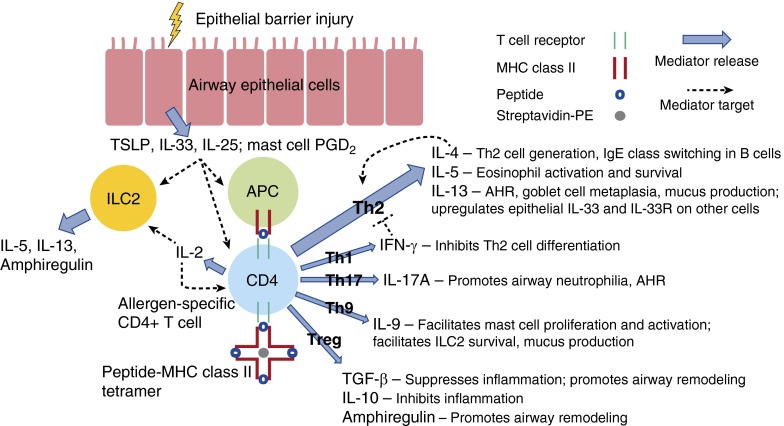
In allergic asthma, aeroallergen exposure of sensitized individuals mobilizes robust innate and adaptive airway immune responses, stimulating eosinophilic airway inflammation and the activation and infiltration of allergen-specific CD4(+) T cells into the airways. Allergen-specific CD4(+) T cells are thought to be central players in the asthmatic response as they specifically recognize the allergen and initiate and orchestrate the asthmatic inflammatory response. In this article, we briefly review the role of allergen-specific CD4(+) T cells in the pathogenesis of human allergic airway inflammation in allergic individuals, discuss the use of allergen-major histocompatibility complex class II tetramers to characterize allergen-specific CD4(+) T cells, and highlight current gaps in knowledge and directions for future research pertaining to the role of allergen-specific CD4(+) T cells in human asthma.

Keywords: CD4-positive T lymphocytes; allergens; helper T type 2 cells; humans; inflammation.


Figures

Figure 1.
Figure 1.
Allergen-specific CD4+ T-cell subsets and their interactions with structural, innate, and regulatory cells in the asthmatic airway. In response to epithelial barrier injury, airway epithelial cells release innate type 2 cytokines, including thymic stromal lymphopoietin (TSLP), IL-33, and/or IL-25, which promotes allergic airway inflammation by acting on helper T type 2 (Th2) cells, type 2 innate lymphoid cells (ILC2s), and antigen-presenting cells (APCs), including dendritic cells (DCs) and macrophages. Activated mast cells release numerous mediators, including prostaglandin D2 (PGD2), which promotes airway hyperresponsiveness (AHR) and facilitates the migration and activation of Th2 cells, ILC2s, and other cells. Allergen that breaches the damaged epithelium can be processed and presented by APCs to prime allergen-specific CD4+ T cells. Depending on the cytokine milieu, allergen-specific CD4+ T cells may become Th2-polarized or may proceed down other differentiation pathways: Th1, Th17, Th9, or regulatory T cell (Treg). Allergen-specific CD4+ T cells can be identified with peptide–MHC class II tetramers, which consist of four MHC class II molecules associated with an antigenic peptide bound to streptavidin linked to phycoerythrin (PE). Activated allergen-specific CD4+ T cells produce IL-2, which acts as a growth factor for T cells and ILC2s. Th2 cells specifically produce IL-4, which promotes the generation of more Th2 cells in a positive feedback loop and class switching in B cells to produce allergen-specific IgE, IL-5, which stimulates eosinophil activation and survival, and IL-13, which promotes AHR, goblet cell metaplasia, mucus production, and may also up-regulate epithelial IL-33 and IL-33 receptor (IL-33R) on other cells. Th1 cells produce IFN-γ, which can counteract Th2-cell differentiation. Th17 cells produce IL-17A, which promotes the production of cytokines and chemokines that recruit neutrophils to the airway and may also promote AHR by enhancing smooth muscle cell contractility. Th9 cells produce IL-9, which facilitates mast cell proliferation and activation, facilitates ILC2 survival, and may also promote mucus production and airway remodeling. Tregs produce IL-10, which can dampen inflammation, and transforming growth factor (TGF)-β, which can suppress inflammation and, along with ILC2-derived amphiregulin, may promote airway remodeling. Impaired regulatory responses may exacerbate the development and persistence of type 2 inflammation. Thick blue arrows indicate the production of a cytokine or mediator by a particular cell; thin dotted black lines indicate the activity of a cytokine or mediator on a particular cell.

References

    1. Mosmann TR, Cherwinski H, Bond MW, Giedlin MA, Coffman RL. Two types of murine helper T cell clone. I. Definition according to profiles of lymphokine activities and secreted proteins. J Immunol. 1986;136:2348–2357. - PubMed
    1. Walker C, Bode E, Boer L, Hansel TT, Blaser K, Virchow JC., Jr Allergic and nonallergic asthmatics have distinct patterns of T-cell activation and cytokine production in peripheral blood and bronchoalveolar lavage. Am Rev Respir Dis. 1992;146:109–115. - PubMed
    1. Robinson DS, Hamid Q, Ying S, Tsicopoulos A, Barkans J, Bentley AM, Corrigan C, Durham SR, Kay AB. Predominant TH2-like bronchoalveolar T-lymphocyte population in atopic asthma. N Engl J Med. 1992;326:298–304. - PubMed
    1. Prescott SL, Macaubas C, Smallacombe T, Holt BJ, Sly PD, Holt PG. Development of allergen-specific T-cell memory in atopic and normal children. Lancet. 1999;353:196–200. - PubMed
    1. Jackson DJ, Makrinioti H, Rana BM, Shamji BW, Trujillo-Torralbo MB, Footitt J, Jerico Del-Rosario, Telcian AG, Nikonova A, Zhu J, et al. IL-33–dependent type 2 inflammation during rhinovirus-induced asthma exacerbations in vivo. Am J Respir Crit Care Med. 2014;190:1373–1382. - PMC - PubMed