. 2017 Aug;25(7):513-523.
doi: 10.1097/PAI.0000000000000360.

Detecting Gene Rearrangements in Patient Populations Through a 2-Step Diagnostic Test Comprised of Rapid IHC Enrichment Followed by Sensitive Next-Generation Sequencing

Affiliations

Detecting Gene Rearrangements in Patient Populations Through a 2-Step Diagnostic Test Comprised of Rapid IHC Enrichment Followed by Sensitive Next-Generation Sequencing

Danielle A Murphy et al. Appl Immunohistochem Mol Morphol. 2017 Aug.

Abstract

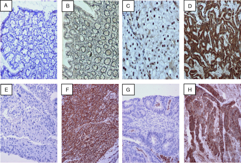
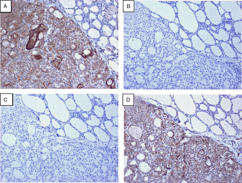
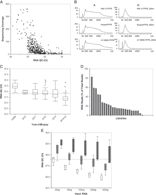
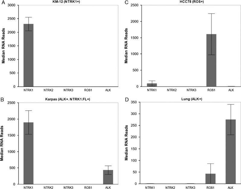
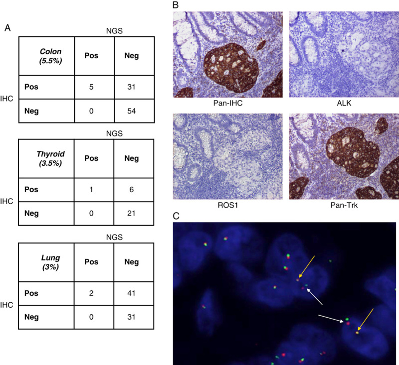
Targeted therapy combined with companion diagnostics has led to the advancement of next-generation sequencing (NGS) for detection of molecular alterations. However, using a diagnostic test to identify patient populations with low prevalence molecular alterations, such as gene rearrangements, poses efficiency, and cost challenges. To address this, we have developed a 2-step diagnostic test to identify NTRK1, NTRK2, NTRK3, ROS1, and ALK rearrangements in formalin-fixed paraffin-embedded clinical specimens. This test is comprised of immunohistochemistry screening using a pan-receptor tyrosine kinase cocktail of antibodies to identify samples expressing TrkA (encoded by NTRK1), TrkB (encoded by NTRK2), TrkC (encoded by NTRK3), ROS1, and ALK followed by an RNA-based anchored multiplex polymerase chain reaction NGS assay. We demonstrate that the NGS assay is accurate and reproducible in identification of gene rearrangements. Furthermore, implementation of an RNA quality control metric to assess the presence of amplifiable nucleic acid input material enables a measure of confidence when an NGS result is negative for gene rearrangements. Finally, we demonstrate that performing a pan-receptor tyrosine kinase immunohistochemistry staining enriches detection of the patient population for gene rearrangements from 4% to 9% and has a 100% negative predictive value. Together, this 2-step assay is an efficient method for detection of gene rearrangements in both clinical testing and studies of archival formalin-fixed paraffin-embedded specimens.


Conflict of interest statement

The authors declare no conflict of interest.

Figures

FIGURE 1
FIGURE 1
Examples of the pan-RTK IHC staining for TrkA, TrkB, TrkC, ROS1, and ALK in multiple tissue types. Tissues were stained with the pan-RTK antibody cocktail and developed with 3,3′-diaminobenzidine (DAB) to detect protein expressions. A and B, Staining of pan-RTK IHC in normal colon (4× magnification). C and D, Staining of pan-RTK in normal adjacent liver and metastatic lung tissue from the same patient on the same slide (respectively, 20× magnification). E and F, Negative and positive staining in lung adenocarcinoma tissue (respectively, 10× magnification). G and H, Negative and positive staining of colon carcinoma tissue (respectively, 10× magnification). IHC indicates immunohistochemistry; RTK, receptor tyrosine kinase.
FIGURE 2
FIGURE 2
Specific pan-RTK IHC staining observed in a thyroid carcinoma confirmed by NGS to harbor an ETV6:NTRK3 fusion. Serial sections from this sample were sequentially stained with the pan-RTK antibody cocktail (A), or with individual antibodies against ALK (B), ROS1 (C), or pan-TRK (TrkA, TrkB, TrkC) (D) and developed with DAB to detect protein expression for these targets. All images were taken at 10× magnification. IHC indicates immunohistochemistry; NGS, next-generation sequencing; RTK, receptor tyrosine kinase.
FIGURE 3
FIGURE 3
Assessment of RNA quality in FFPE tissues. A, FFPE tissues (n=478) were subjected to the RNA QC assay followed by NGS. Quantitative values for the RNA QC assay (Ct value) and sequencing coverage obtained from NGS (unique RNA reads) were graphed to examine the correlation between quality of RNA and the ability of the RNA to generate sequence. B, Three different FFPE-embedded cell lines harboring gene rearrangements (KM12; TPM3:NTRK1, KARPAS-299; NPM1:ALK, and U-118 MG; GOPC:ROS1) were mechanically sheared by Covaris to generate RNA fragments that range in average size from ∼300 nt (short) to ∼1000 nt (long). The electropherogram image shows the RNA profiles of each sample: A=normal (left column) and A′=sheared (right column). C, RNA obtained from FFPE blocks with increasing years from biopsy date (ranging from <2009 to 2015) was investigated for quality by running the RNA QC assay and assessing the Ct value obtained (n>6 for each year, circle indicates outliers, P<0.0001 for linear trend). D, Libraries were prepared from FFPE samples (n=25; different tissue types) using the same RNA input amount for each library (200 ng). The libraries were then sequenced by NGS and analyzed by Archer Analysis for the RNA reads per sample. E, To determine how quality of RNA impacts input amount, we assessed the RNA Ct values of FFPE samples when varying input amounts. Samples were stratified based on Ct values at 200 ng input (n≥6 for each category). White=low, Ct<26, diagonal hashes=medium, Ct=26 to 29, and checkered=high, Ct>29. The dotted line represents the threshold at 29. Ct indicates cycle threshold; FFPE, formalin-fixed paraffin-embedded; NGS, next-generation sequencing; QC, quality control.
FIGURE 4
FIGURE 4
FFPE-embedded cell lines with known gene rearrangements in NTRK1 (KM-12, A), ALK with overexpression of full length NTRK1 (NTRK1 FL+) (KARPAS-299, B), ROS1 (HCC78, C), and a lung adenocarcinoma tumor with an EML4:ALK gene rearrangement (D) were run in triplicate using the NGS assay. Samples were then examined for median unique RNA reads for each gene rearrangement receptor (NTRK1, NTRK2, NTRK3, ROS1, and ALK). FFPE indicates formalin-fixed paraffin-embedded; NGS, next-generation sequencing.
FIGURE 5
FIGURE 5
Clinical relevance and results of using pan-RTK IHC screening followed by NGS to detect gene rearrangements. A, Clinical FFPE samples (colon, thyroid, and lung cancer) were stained with a pan-RTK antibody cocktail; IHC was performed and samples were qualitatively scored as negative or positive for expression of the proteins. RNA extracted from these samples was run on the NGS assay and qualitatively assessed for detection of gene rearrangements. IHC and NGS data were correlated for positive and negative agreement between protein expression by IHC and detection of gene rearrangement by NGS. B, Example of a clinical patient sample harboring a TPM3:NTRK1 gene rearrangement detected by the NGS assay and run through the pan-RTK IHC (top left panel) as well as individual IHC stains for ALK (top right panel), ROS1 (bottom left panel), and Pan-Trk (bottom right panel). All images were taken at 10× magnification. C, The clinical patient sample in (B) was stained by FISH using NTRK1 break-apart gene rearrangement probes and examined for NTRK1 gene rearrangement positivity. White arrows indicate NTRK1 fusion positive cells and yellow arrows indicate wild-type NTRK1 expression. FFPE indicates formalin-fixed paraffin-embedded; FISH, fluorescence in-situ hybridization; IHC, immunohistochemistry; NGS, next-generation sequencing; RTK, receptor tyrosine kinase.

References

    1. Shaw AT, Hsu PP, Awad MM, et al. Tyrosine kinase gene rearrangements in epithelial malignancies. Nat Rev Cancer. 2013;13:772–787. - PMC - PubMed
    1. Shaw AT, Ou Sai-Hong I, Bang Yung-Jue, et al. Crizotinib in ROS1—rearranged non–small-cell lung cancer. N Engl J Med. 2014;371:1963–1971. - PMC - PubMed
    1. Kwak EL, Bang Yung-Jue, Camidge D Ross, et al. Anaplastic lymphoma kinase inhibition in non-small-cell lung cancer. N Engl J Med. 2010;363:1693–1703. - PMC - PubMed
    1. Bergethon K, Shaw Alice T, Ignatius Ou Sai-Hong, et al. ROS1 rearrangements define a unique molecular class of lung cancers. J Clin Oncol. 2012;30:863–870. - PMC - PubMed
    1. Davies KD, Le Anh T, Theodoro Mariana F, et al. Identifying and targeting ROS1 gene fusions in non-small cell lung cancer. Clin Cancer Res. 2012;18:4570–4579. - PMC - PubMed