

Prognostic value and clinicopathologic characteristics of L1 cell adhesion molecule (L1CAM) in a large series of vulvar squamous cell carcinomas
- PMID: 27028855
- PMCID: PMC5041974
- DOI: 10.18632/oncotarget.8353
Prognostic value and clinicopathologic characteristics of L1 cell adhesion molecule (L1CAM) in a large series of vulvar squamous cell carcinomas
Abstract
Background: Vulvar cancer treatment is mostly curative, but also has high morbidity rates. In a search for markers that can identify patients at risk of metastases, we investigated the prognostic value of L1-cell adhesion molecule (L1CAM) in large series of vulvar squamous cell carcinomas (VSCCs). L1CAM promotes cell motility and is an emerging prognostic factor for metastasis in many cancer subtypes.
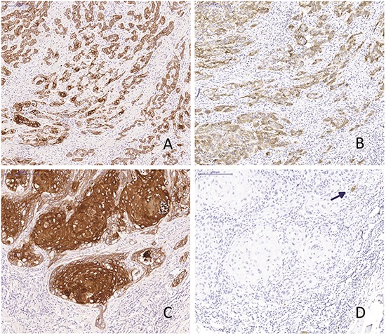
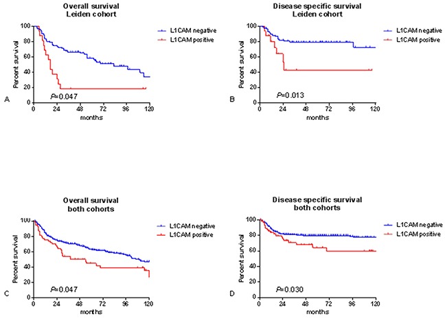
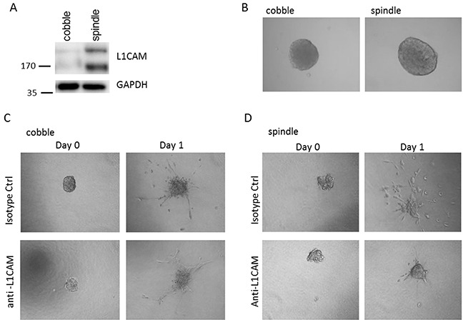
Results: L1CAM expression was observed at the invasive front or in spray-patterned parts of 17% of the tumours. L1CAM-positive tumours expressed vimentin more often, but L1CAM expression was not associated with TP53 or CTNNB1 mutations. Five-year survival was worse for patients with L1CAM expression (overall survival 46.1% vs 63.6%, P=.014, disease specific survival 63.8% vs 80.0%, P=.018). Multivariate analysis indicates L1CAM expression as an independent prognostic marker (HR 2.9, 95% CI 1.10-7.68). An in vitro spheroid invasion assay showed decreased invasion of L1CAM-expressing VSCC spindle cells after treatment with L1CAM-neutralising antibodies.
Methods: Paraffin-embedded tumour tissue from two cohorts (N=103 and 245) of primary VSCCs were stained for L1CAM, vimentin and E-cadherin. Patients of the first cohort were tested for human papilloma virus infection and sequenced for TP53 and CTNNB1 (β-catenin) mutations. The expression of L1CAM was correlated to clinical characteristics and patient survival.
Conclusion: This is the first study to show high L1CAM-expression at the infiltrating margin of VSCC's. L1CAM-expressing VSCCs had a significantly worse prognosis compared to L1CAM-negative tumours. The highest expression was observed in spindle-shaped cells, where it might be correlated to their invasive capacity.
Keywords: L1 cell adhesion molecule; L1CAM; squamous cell carcinoma; survival; vulvar cancer.
Conflict of interest statement
The authors declare no conflicts of interest.
Figures
Similar articles
-
L1 cell adhesion molecule (L1CAM) in stage IB cervical cancer: distinct expression in squamous cell carcinomas and adenocarcinomas.J Clin Pathol. 2020 Nov;73(11):748-753. doi: 10.1136/jclinpath-2020-206500. Epub 2020 May 4. J Clin Pathol. 2020. PMID: 32366597
-
L1CAM protein expression is associated with poor prognosis in non-small cell lung cancer.Mol Cancer. 2011 Oct 10;10:127. doi: 10.1186/1476-4598-10-127. Mol Cancer. 2011. PMID: 21985405 Free PMC article.
-
L1 cell adhesion molecule is a strong predictor for distant recurrence and overall survival in early stage endometrial cancer: pooled PORTEC trial results.Eur J Cancer. 2014 Oct;50(15):2602-10. doi: 10.1016/j.ejca.2014.07.014. Epub 2014 Aug 7. Eur J Cancer. 2014. PMID: 25126672
-
L1CAM in human cancer.Int J Cancer. 2016 Apr 1;138(7):1565-76. doi: 10.1002/ijc.29658. Epub 2015 Aug 25. Int J Cancer. 2016. PMID: 26111503 Review.
-
High L1CAM expression predicts poor prognosis of patients with endometrial cancer: A systematic review and meta-analysis.Medicine (Baltimore). 2021 Apr 2;100(13):e25330. doi: 10.1097/MD.0000000000025330. Medicine (Baltimore). 2021. PMID: 33787629 Free PMC article.
Cited by
-
Prognostic significance of L1 cell adhesion molecule in cancer patients: A systematic review and meta-analysis.Oncotarget. 2016 Dec 20;7(51):85196-85207. doi: 10.18632/oncotarget.13236. Oncotarget. 2016. PMID: 27833079 Free PMC article.
-
Mint3-mediated L1CAM expression in fibroblasts promotes cancer cell proliferation via integrin α5β1 and tumour growth.Oncogenesis. 2017 May 15;6(5):e334. doi: 10.1038/oncsis.2017.27. Oncogenesis. 2017. PMID: 28504692 Free PMC article.
-
Endoglin and squamous cell carcinomas.Front Med (Lausanne). 2023 Jun 16;10:1112573. doi: 10.3389/fmed.2023.1112573. eCollection 2023. Front Med (Lausanne). 2023. PMID: 37396898 Free PMC article.
-
L1 cell adhesion molecule (L1CAM) is a strong predictor for locoregional recurrences in cervical cancer.Oncotarget. 2017 Sep 18;8(50):87568-87581. doi: 10.18632/oncotarget.20976. eCollection 2017 Oct 20. Oncotarget. 2017. PMID: 29152102 Free PMC article.
References
-
- Dittmer C, Fischer D, Diedrich K, Thill M. Diagnosis and treatment options of vulvar cancer: a review. Arch Gynecol Obstet. 2012;285:183–93. - PubMed
-
- Judson PL, Habermann EB, Baxter NN, Durham SB, Virnig BA. Trends in the incidence of invasive and in situ vulvar carcinoma. Obstet Gyneco.l. 2006;107:1018–22. - PubMed
-
- Hacker NF. Vulvar Cancer. In: Berek JS, Hacker NF, editors. Practical gynecologic oncology. 3. Philadelphia: Lippincott Williams & Wilkins; 2005. pp. 543–76.
-
- van de Nieuwenhof HP, Massuger LF, van der Avoort IA, Bekkers RL, Casparie M, Abma W, van Kempen LC, de Hullu JA. Vulvar squamous cell carcinoma development after diagnosis of VIN increases with age. Eur J Cancer. 2009;45:851–6. - PubMed
-
- Del Pino M, Rodriguez-Carunchio L, Ordi J. Pathways of vulvar intraepithelial neoplasia and squamous cell carcinoma. Histopathology. 2013;62:161–75. - PubMed
MeSH terms
Substances
LinkOut - more resources
Full Text Sources
Other Literature Sources
Medical
Research Materials
Miscellaneous
