. 2016 May 20;291(21):11285-99.
doi: 10.1074/jbc.M115.710681. Epub 2016 Mar 30.

Protein Kinase D and Gβγ Subunits Mediate Agonist-evoked Translocation of Protease-activated Receptor-2 from the Golgi Apparatus to the Plasma Membrane

Affiliations

Protein Kinase D and Gβγ Subunits Mediate Agonist-evoked Translocation of Protease-activated Receptor-2 from the Golgi Apparatus to the Plasma Membrane

Dane D Jensen et al. J Biol Chem. .

Abstract

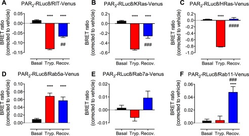
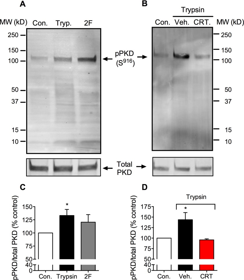
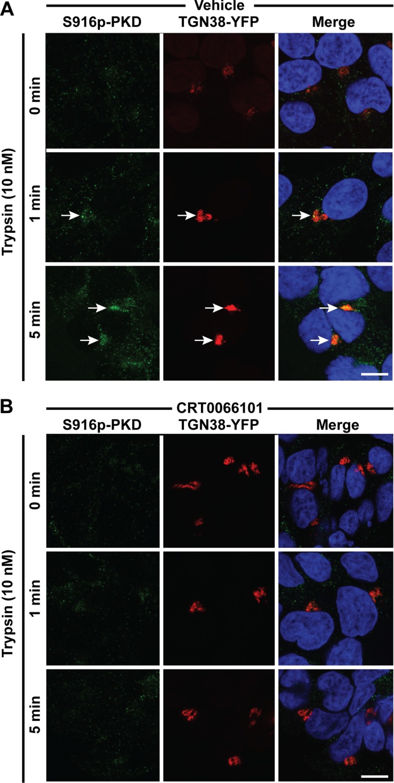
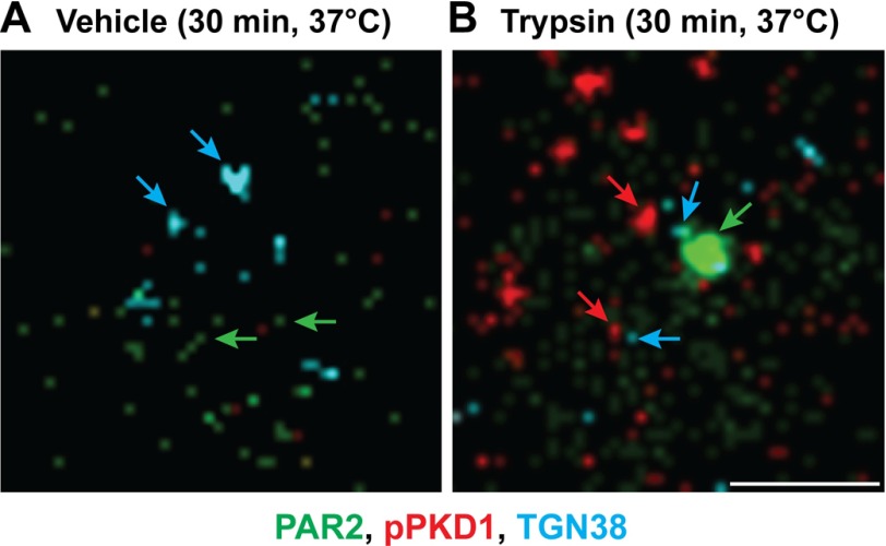
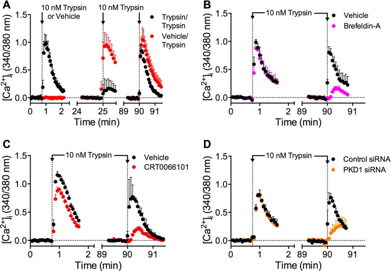
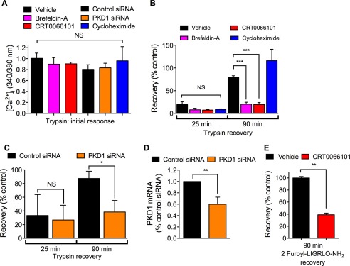
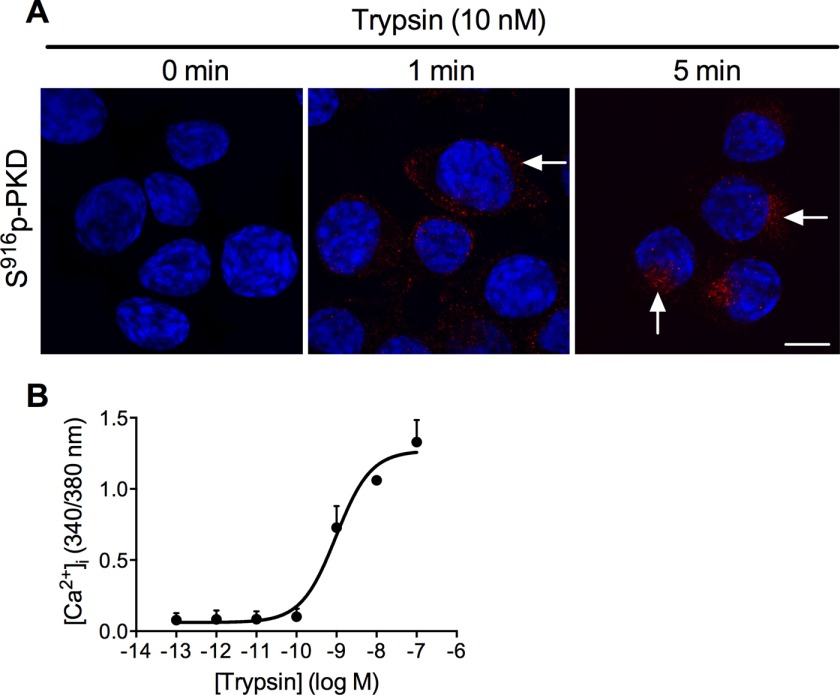
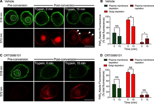
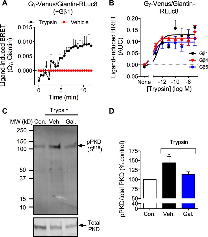
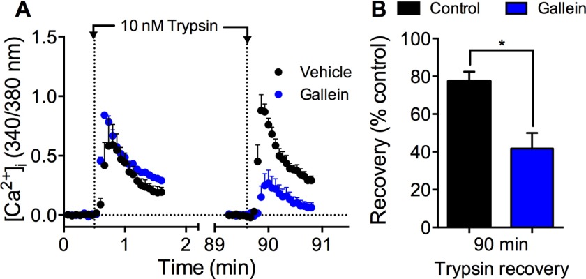
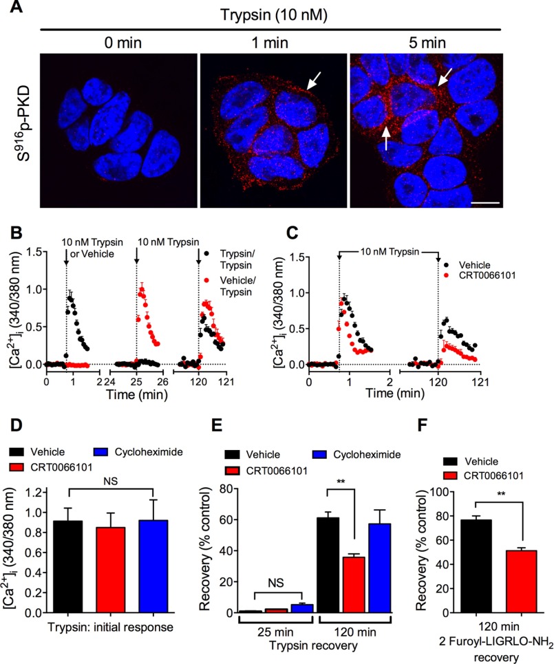
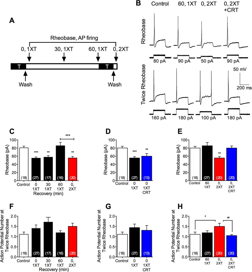
Agonist-evoked endocytosis of G protein-coupled receptors has been extensively studied. The mechanisms by which agonists stimulate mobilization and plasma membrane translocation of G protein-coupled receptors from intracellular stores are unexplored. Protease-activated receptor-2 (PAR2) traffics to lysosomes, and sustained protease signaling requires mobilization and plasma membrane trafficking of PAR2 from Golgi stores. We evaluated the contribution of protein kinase D (PKD) and Gβγ to this process. In HEK293 and KNRK cells, the PAR2 agonists trypsin and 2-furoyl-LIGRLO-NH2 activated PKD in the Golgi apparatus, where PKD regulates protein trafficking. PAR2 activation induced translocation of Gβγ, a PKD activator, to the Golgi apparatus, determined by bioluminescence resonance energy transfer between Gγ-Venus and giantin-Rluc8. Inhibitors of PKD (CRT0066101) and Gβγ (gallein) prevented PAR2-stimulated activation of PKD. CRT0066101, PKD1 siRNA, and gallein all inhibited recovery of PAR2-evoked Ca(2+) signaling. PAR2 with a photoconvertible Kaede tag was expressed in KNRK cells to examine receptor translocation from the Golgi apparatus to the plasma membrane. Irradiation of the Golgi region (405 nm) induced green-red photo-conversion of PAR2-Kaede. Trypsin depleted PAR2-Kaede from the Golgi apparatus and repleted PAR2-Kaede at the plasma membrane. CRT0066101 inhibited PAR2-Kaede translocation to the plasma membrane. CRT0066101 also inhibited sustained protease signaling to colonocytes and nociceptive neurons that naturally express PAR2 and mediate protease-evoked inflammation and nociception. Our results reveal a major role for PKD and Gβγ in agonist-evoked mobilization of intracellular PAR2 stores that is required for sustained signaling by extracellular proteases.

Keywords: 7-helix receptor; Golgi; protein kinase D (PKD); protein trafficking (Golgi); receptor desensitization.


Figures

FIGURE 1.
FIGURE 1.
Trypsin-induced PAR2 endocytosis and recovery. HEK293 cells expressing PAR2-RLuc8 and RIT (A), KRas (B), HRas (C), Rab5a (D), Rab7a (E), or Rab11 (F) tagged with Venus were stimulated with trypsin (100 nm, 20 min). Cells were washed and recovered in trypsin-free medium for 2 h. BRET was measured after trypsin (Tryp.) and after recovery (Recov.). ****, p < 0.0001 compared with basal; ##, p < 0.01; ###, p < 0.001; ####, p < 0.0001 compare with trypsin, n = 3 experiments.
FIGURE 2.
FIGURE 2.
PAR2-induced activation of PKD. Western blots for Ser916 phospho-PKD (p-PKD) and total PKD in HEK293 cell are shown. Cells were unstimulated (control, Con.) or were stimulated with trypsin (Tryp., 10 nm) or 2-furoyl-LIGRLO-NH2 (2F, 10 μm) for 5 min. Cells were preincubated with CRT0066101 (CRT) or vehicle (Veh.). A and B, representative blots. C and D, quantified results. *, p < 0.05, n = 3 experiments.
FIGURE 3.
FIGURE 3.
Trypsin-induced activation of PKD in the Golgi apparatus. HEK293 cells expressing TGN38-YFP were stimulated with trypsin (10 nm, 0, 1, or 5 min). Ser916 phospho-PKD was detected by immunofluorescence and confocal microscopy. A, vehicle control. B, CRT0066101-treated cells. Scale, 10 μm.
FIGURE 4.
FIGURE 4.
Trypsin-induced localization of phospho-PKD in proximity to PAR2 in the Golgi apparatus. HEK-FLAG-PAR2-HA cells were incubated with vehicle (A) or trypsin (10 nm) (B) for 30 min at 37 °C. Cells were fixed, and Ser916 phospho-PKD (pPKD, red), PAR2 (green) and TGN38 (blue) were localized by immunofluorescence and super-resolution microscopy. Representative images from >3 experiments are shown. Scale, 1 μm.
FIGURE 5.
FIGURE 5.
PKD-mediated recovery of trypsin-evoked Ca2+ signaling. HEK293 cells were challenged with vehicle or trypsin (10 nm, 10 min), washed, and challenged again with trypsin (10 nm) 25 or 90 min later. [Ca2+]i was measured. A, time course showing desensitization at 25 min and recovery at 90 min. B–D, effects of brefeldin-A (B), CRT0066101 (C), or control siRNA or PKD1 siRNA (D) on recovery at 90 min. n = 3 experiments.
FIGURE 6.
FIGURE 6.
PKD-mediated recovery of trypsin-evoked Ca2+ signaling. A–C and E, HEK293 cells were preincubated with vehicle, brefeldin-A, CRT0066101, or cycloheximide or were transfected with control siRNA or PKD1-siRNA. Cells were challenged with vehicle, trypsin (10 nm, 10 min), or 2-furoyl-LIGRLO-NH2 (10 μm, 10 min), washed, and challenged again with trypsin (10 nm) or 2-furoyl-LIGRLO-NH2 (10 μm) 25 or 90 min later. [Ca2+]i was measured. A, initial response to trypsin. B, recovery of trypsin responses at 25 and 90 min. C, effects of control siRNA or PKD1 siRNA on recovery of trypsin responses at 25 and 90 min. D, PKD expression at 24 h after transfection with control siRNA or PKD1 siRNA. E, recovery of 2-furoyl-LIGRLO-NH2 response at 90 min. *, p < 0.05; **, p < 0.01; ***, p < 0.0001, NS, not significant, n = 3 experiments.
FIGURE 7.
FIGURE 7.
Trypsin-induced activation of PKD and PAR2 in KNRK-PAR2-Kaede cells. A, localization of Ser916 phospho-PKD in KNRK-PAR2-Kaede cells treated with trypsin (10 nm, 0, 1, and 5 min). Scale, 10 μm. B, trypsin-evoked changes in [Ca2+]i in KNRK-PAR2-Kaede cells. n = 3 experiments.
FIGURE 8.
FIGURE 8.
PKD-mediated plasma membrane translocation of PAR2-Kaede. PAR2-Kaede in the vicinity of the Golgi apparatus of KNRK-PAR2-Kaede cells was green-red photoconverted using a confocal laser (illuminated region: dashed circle). Cells were incubated with trypsin (10 nm) for 0 or 15 min. PAR2-Kaede fluorescence was measured at 518 and 572 nm at the plasma membrane and in the vicinity of the Golgi apparatus at 0 and 15 min. A and C, representative images of cells treated with vehicle (A) or CRT0066101 (C). Scale, 10 μm. B and D, quantification of plasma membrane depletion of non-converted PAR2-Kaede (518 nm, green), Golgi depletion of converted PAR2-Kaede (572 nm, red), and plasma membrane repletion of PAR2-Kaede (572 nm, red). *, p < 0.05; **, p < 0.001, NS, not significant, n = 5 experiments, >45 cells analyzed per condition.
FIGURE 9.
FIGURE 9.
Gβγ translocation to the Golgi apparatus and activation of PKD. A and B, trypsin-induced BRET between Gγ-Venus and giatin-RLuc8 in HEK293 cells coexpressing Gβ1, Gβ4, or Gβ5. A, time course trypsin response. B, trypsin concentration-response, area under curve (AUC) over 15 min. n = 4–7 experiments. C and D, Western blot for Ser916 phospho-PKD and total PKD in HEK cells. Cells were unstimulated (control, Con.) or were stimulated with trypsin (10 nm) for 5 min. Some cells were preincubated with gallein (Gal) or vehicle (Veh.). C, representative blot. D, quantified results. *, p < 0.05, n = 3 experiments.
FIGURE 10.
FIGURE 10.
Gβγ-mediated recovery of trypsin-evoked Ca2+ signaling. HEK293 cells were challenged with vehicle or trypsin (10 nm, 5 min), washed, and challenged again with trypsin (10 nm) 90 min later. [Ca2+]i was measured. A, effects of gallein or vehicle on kinetics. B, effects of gallein on recovery at 90 min. *, p < 0.05, n = 3 experiments.
FIGURE 11.
FIGURE 11.
Trypsin-induced activation of PKD and PKD-mediated recovery of trypsin-evoked Ca2+ signaling in colonocytes. A, localization of Ser916 phospho-PKD in NCM460 cells treated with trypsin (10 nm, 0, 1, and 5 min). Scale, 10 μm. B–E, NCM460 cells were preincubated with vehicle or CRT0066101. Cells were challenged with vehicle, trypsin (10 nm, 10 min), or 2-furoyl-LIGRLO-NH2 (10 μm, 10 min), washed, and challenged again with trypsin (10 nm) or 2-furoyl-LIGRLO-NH2 (10 μm) 25 or 120 min later. [Ca2+]i was measured. B, time course showing desensitization at 25 min and recovery at 120 min. C, effects of CRT0066101 on recovery of trypsin responses at 120 min. D, initial response to trypsin. E, recovery of trypsin responses at 25 and 120 min. F, recovery of 2-furoyl-LIGRLO-NH2 response at 120 min. **, p < 0.001. NS, not significant, n = 3 experiments.
FIGURE 12.
FIGURE 12.
PKD-mediated sustained neuronal hyperexcitability. A, dorsal root ganglion neurons were exposed to trypsin (T, 50 nm) for 10 min, washed, and incubated in trypsin-free medium. Excitability was assessed at 0 (0, 1XT), 30 (30, 1XT), or 60 (60, 1XT) min after first trypsin (0, 1XT). Some neurons were subjected to a second trypsin exposure 60 min after the first and were washed, and excitability was assessed (0, 2XT). B, representative action potential firing at rheobase and twice rheobase. C and D, pooled measurements of rheobase. F–H, pooled measurements of action potential firing at twice rheobase. *, p < 0.05; **, p < 0.001; ***, p < 0.0001 compared with control; ##, p < 0.001; ###, p < 0.0001. n denotes number of recorded neurons from studies of 16 mice.

Similar articles

Cited by

References

    1. Kang D. S., Tian X., and Benovic J. L. (2014) Role of β-arrestins and arrestin domain-containing proteins in G protein-coupled receptor trafficking. Curr. Opin. Cell Biol. 27, 63–71 - PMC - PubMed
    1. McConalogue K., Déry O., Lovett M., Wong H., Walsh J. H., Grady E. F., and Bunnett N. W. (1999) Substance P-induced trafficking of β-arrestins. The role of β-arrestins in endocytosis of the neurokinin-1 receptor. J. Biol. Chem. 274, 16257–16268 - PubMed
    1. Williams J. T., Ingram S. L., Henderson G., Chavkin C., von Zastrow M., Schulz S., Koch T., Evans C. J., and Christie M. J. (2013) Regulation of μ-opioid receptors: desensitization, phosphorylation, internalization, and tolerance. Pharmacol. Rev. 65, 223–254 - PMC - PubMed
    1. Déry O., Thoma M. S., Wong H., Grady E. F., and Bunnett N. W. (1999) Trafficking of proteinase-activated receptor-2 and β-arrestin-1 tagged with green fluorescent protein. β-Arrestin-dependent endocytosis of a proteinase receptor. J. Biol. Chem. 274, 18524–18535 - PubMed
    1. Oh P., Horner T., Witkiewicz H., and Schnitzer J. E. (2012) Endothelin induces rapid, dynamin-mediated budding of endothelial caveolae rich in ET-B. J. Biol. Chem. 287, 17353–17362 - PMC - PubMed

Publication types

MeSH terms