. 2016 May 27;90(12):5622-5635.
doi: 10.1128/JVI.00446-16. Print 2016 Jun 15.

Roles of Us8A and Its Phosphorylation Mediated by Us3 in Herpes Simplex Virus 1 Pathogenesis

Affiliations

Roles of Us8A and Its Phosphorylation Mediated by Us3 in Herpes Simplex Virus 1 Pathogenesis

Akihisa Kato et al. J Virol. .

Abstract

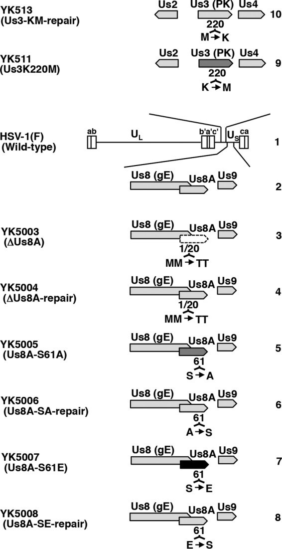
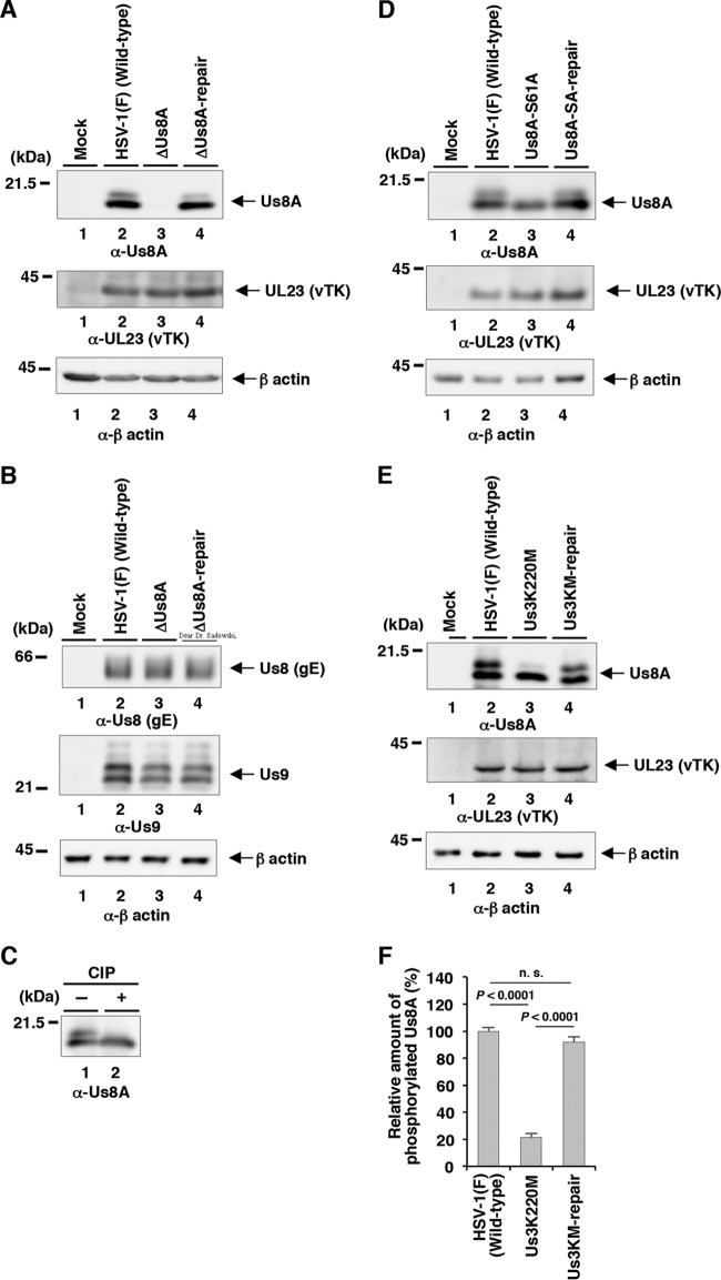
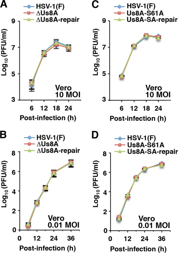
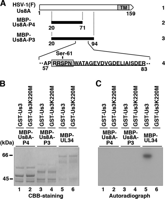
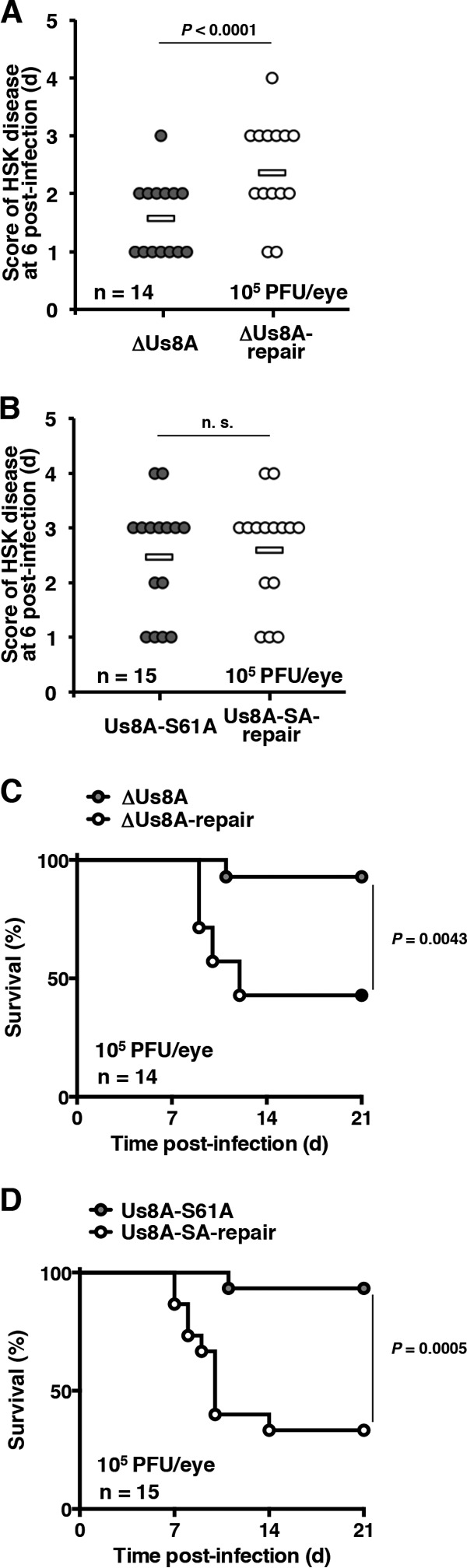
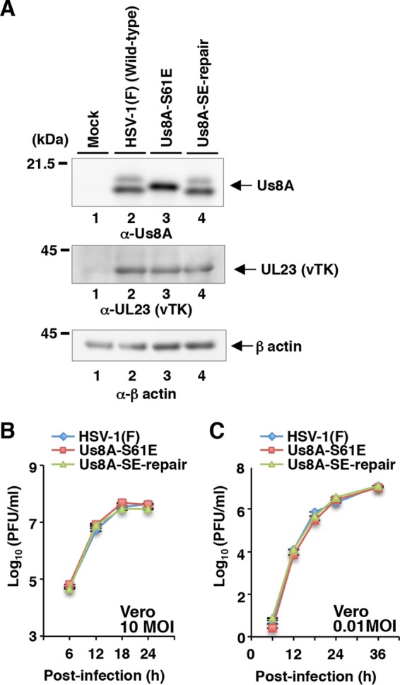
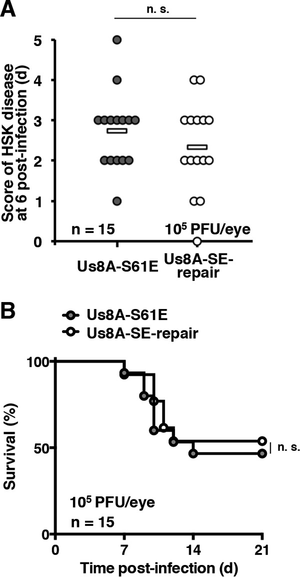
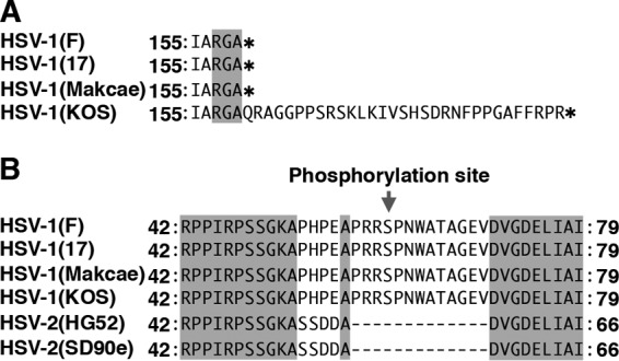
The herpes simplex virus 1 (HSV-1) Us8A gene overlaps the gene that encodes glycoprotein E (gE). Previous studies have investigated the roles of Us8A in HSV-1 infection using null mutations in Us8A and gE; therefore, the role of Us8A remains to be elucidated. In this study, we investigated the function of Us8A and its phosphorylation at serine 61 (Ser-61), which we recently identified as a phosphorylation site by mass spectrometry-based phosphoproteomic analysis of HSV-1-infected cells, in HSV-1 pathogenesis. We observed that (i) the phosphorylation of Us8A Ser-61 in infected cells was dependent on the activity of the virus-encoded Us3 protein kinase; (ii) the Us8A null mutant virus exhibited a 10-fold increase in the 50% lethal dose for virulence in the central nervous system (CNS) of mice following intracranial infection compared with a repaired virus; (iii) replacement of Ser-61 with alanine (S61A) in Us8A had little effect on virulence in the CNS of mice following intracranial infection, whereas it significantly reduced the mortality of mice following ocular infection to levels similar to the Us8A null mutant virus; (iv) the Us8A S61A mutation also significantly reduced viral yields in mice following ocular infection, mainly in the trigeminal ganglia and brains; and (v) a phosphomimetic mutation at Us8A Ser-61 restored wild-type viral yields and virulence. Collectively, these results indicate that Us8A is a novel HSV-1 virulence factor and suggest that the Us3-mediated phosphorylation of Us8A Ser-61 regulates Us8A function for viral invasion into the CNS from peripheral sites.

Importance: The DNA genomes of viruses within the subfamily Alphaherpesvirinae are divided into unique long (UL) and unique short (Us) regions. Us regions contain alphaherpesvirus-specific genes. Recently, high-throughput sequencing of ocular isolates of HSV-1 showed that Us8A was the most highly conserved of 13 herpes simplex virus 1 (HSV-1) genes mapped to the Us region, suggesting Us8A may have an important role in the HSV-1 life cycle. However, the specific role of Us8A in HSV-1 infection remains to be elucidated. Here, we show that Us8A is a virulence factor for HSV-1 infection in mice, and the function of Us8A for viral invasion into the central nervous system from peripheral sites is regulated by Us3-mediated phosphorylation of the protein at Ser-61. This is the first study to report the significance of Us8A and its regulation in HSV-1 infection.


Figures

FIG 1
FIG 1
Strategy to construct a self-excisable HSV-1(F) BAC clone. Shown is a schematic diagram of the genome structure of HSV-1(F), the intergenic regions of recombinant viruses with or without the BAC sequence and the EGFP expression cassette insertions, and E. coli containing HSV-1(F) BAC. Line 1, wild-type HSV-1(F) genome; line 2, intergenic region between HSV-1(F) UL50 and UL51 genes; lines 3 and 7, schematic diagrams of recombinant viruses YK5000 and YK5002, respectively; lines 4 and 5, schematic diagrams of E. coli plasmid pYEbac5001 contained in DH10B and GS1783, respectively; line 6, schematic diagram of E. coli plasmid pYEbac5002 contained in GS1783.
FIG 2
FIG 2
Schematic diagrams of the genome structures of HSV-1(F) and recombinant viruses used in this study. Line 1, wild-type HSV-1(F) genome; line 2, domain carrying the Us8 (gE), Us8A, and Us9 open reading frames; lines 3 to 8, recombinant viruses with mutations in the Us8A gene; lines 9 and 10, recombinant viruses with a mutation in the Us3 gene.
FIG 3
FIG 3
Viral genomes, expression of viral proteins, and growth curves from cells infected with HSV-1(F) and YK5002 (ΔBAC). (A) Agarose gel electrophoresis of BamHI-digested viral DNAs of HSV-1(F) (lane 1) and recombinant virus YK5002 (ΔBAC) (lane 2). Fragment a (5.3 kb) and fragment b (5.2 kb) were detected in the YK5002 and HSV-1(F) genomes, respectively, as a result of insertion of the loxP sequence. (B) Agarose gel electrophoresis of PCR products containing OriS (top) and OriL (bottom) of wild-type HSV-1(F) (lanes 1) and YK5002 (ΔBAC) (lanes 2). (C) Vero cells were mock infected (lanes 1) or infected with wild-type HSV-1(F) (lanes 2) or YK5002 (ΔBAC) (lanes 3) at an MOI of 10, harvested at 18 h postinfection, and then analyzed by immunoblotting with the indicated antibodies. Molecular size (A and B) and mass (C) markers are shown on the left. (D and E) Vero cells were infected at an MOI of 10 (D) or 0.01 (E) with wild-type HSV-1(F) or YK5002 (ΔBAC). Total virus from the cell culture supernatants and in fected cells was harvested at the indicated times and assayed on Vero cells. Each value represents the mean ± standard error of the mean (SEM) of the results of three independent experiments.
FIG 4
FIG 4
Us3-mediated phosphorylation of Us8A Ser-61 in infected cells. (A and B) Vero cells were mock infected (lanes 1) or infected with wild-type HSV-1(F) (lanes 2), YK5003 (ΔUs8A) (lanes 3), or YK5004 (ΔUs8A-repair) (lanes 4) at an MOI of 10; harvested at 18 h postinfection; and then analyzed by immunoblotting with the indicated antibodies. (C) The infected Vero cell lysates were untreated (lane 1) or treated with CIP (lane 2) and then analyzed as described for panel A. (D) Vero cells were mock infected (lanes 1) or infected with wild-type HSV-1(F) (lanes 2), YK5005 (Us8A-S61A) (lanes 3), or YK5006 (Us8A-SA-repair) (lanes 4) at an MOI of 10; harvested at 18 h postinfection; and then analyzed as described for panel A. (E) Vero cells were mock infected (lanes 1) or infected with wild-type HSV-1(F) (lanes 2), YK511 (Us3K220M) (lanes 3), or YK513 (Us3K220M-repair) (lanes 4) at an MOI of 10; harvested at 18 h postinfection; and then analyzed as described for panel A. Molecular mass markers are shown on the left. (F) Quantitation of the amount of the slower-migrating Us8A band relative to that of the faster-migrating Us8A band shown in panel E (top, lanes 2, 3, and 4). Each value represents the mean and SEM of the results of three independent experiments and is expressed relative to the mean value of wild-type HSV-1(F)-infected cells, which was normalized to 100. The statistical significance according to ANOVA and Tukey's test is indicated. n.s., not significant.
FIG 5
FIG 5
Effects of Us8A null or S61A mutations on growth curves for HSV-1-infected cells. Vero cells were infected at an MOI of 10 (A and C) or 0.01 (B and D) with wild-type HSV-1(F), YK5003 (ΔUs8A), or YK5004 (ΔUs8A-repair) (A and B) or wild-type HSV-1(F), YK5005 (Us8A-S61A), or YK5006 (Us8A-SA-repair) (C and D). Total virus from cell culture supernatants and infected cells was harvested at the indicated times and assayed on Vero cells. Each value represents the mean ± SEM of the results of three independent experiments.
FIG 6
FIG 6
Us3-mediated phosphorylation of Us8A in vitro. (A) Schematic diagram of Us8A. Line 1, structure of the Us8A open reading frame. The shaded area represents a predicted transmembrane domain. Line 2, domain of the Us8A gene encoding Us8A residues 20 to 71, which were used in these studies to generate the MBP-Us8A-P4 fusion protein. Line 3, amino acid sequence of Us8A residues 20 to 94. Line 4, domain of the Us8A gene encoding Us8A residues 57 to 83. The amino acid sequence similar to the consensus sequence recognized by Us3 is boxed. A phosphopeptide detected in our previous studies (30, 31) is underlined. (B) Purified MBP-Us8A-P4 (lanes 1 and 2), MBP-Us8A-P3 (lanes 3 and 4), and MBP-UL34 (lanes 5 and 6) were incubated in kinase buffer containing [γ-32P]ATP and purified GST-Us3 (lanes 1, 3, and 5) or GST-Us3K220M (lanes 2, 4, and 6); separated on an SDS-PAGE gel; and stained with CBB. Molecular mass markers are shown on the left. (C) Autoradiograph of the gel in panel B. Molecular mass markers are shown on the left.
FIG 7
FIG 7
Effects of Us8A null and S61A mutations on the development of HSK and mortality of ocularly infected mice. (A and B) Fourteen (A) or 15 (B) 5-week-old female ICR mice infected ocularly with 1 × 10⁵ PFU/eye of YK5003 (ΔUs8A) (A), YK5004 (ΔUs8A-repair) (A), YK5005 (Us8A-S61A) (B), or YK5006 (Us8A-SA-repair) (B) were scored for HSK at 6 days postinfection. Each data point represents the HSK score from one mouse. The horizontal bars represent the mean for each group. The statistical significance according to a two-tailed Student t test is indicated. n.s., not significant. (C and D) Survival of mice in the experiment described for panels A and B was monitored for 21 days postinfection. The statistical significance according to a log rank test is shown.
FIG 8
FIG 8
Effects of the Us8A S61E mutation on expression of Us8A and growth of HSV-1 in infected cells. (A) Vero cells were mock infected (lanes 1) or infected with wild-type HSV-1(F) (lanes 2), YK5007 (Us8A-S61E) (lanes 3), or YK5008 (ΔUs8A-SE-repair) (lanes 4) at an MOI of 10; harvested at 18 h postinfection; and then analyzed by immunoblotting with the indicated antibodies. Molecular mass markers are shown on the left. (B and C) Vero cells were infected at an MOI of 10 (B) or 0.01 (C) with wild-type HSV-1(F), YK5007 (Us8A-S61E), or YK5008 (ΔUs8A-SE-repair). Total virus from the cell culture supernatants and infected cells was harvested at the indicated times and assayed on Vero cells. Each value represents the mean ± SEM of the results of three independent experiments.
FIG 9
FIG 9
Effects of Us8A S61E mutation on the development of HSK and mortality in ocularly infected mice. (A) Fifteen 5-week-old female ICR mice infected ocularly with 1 × 10⁵ PFU/eye of YK5007 (Us8A-S61E) or YK5008 (ΔUs8A-SE-repair) were scored for HSK at 6 days postinfection. Each data point represents the HSK score from one mouse. The horizontal bars represent the mean for each group. n.s., not significant. (B) Survival of mice in the experiment described for panel A was monitored for 21 days postinfection.
FIG 10
FIG 10
Effects of Us8A S61A or S61E mutations on viral yields in the eyes, trigeminal ganglia (TG), and brains of ocularly infected mice. Fifteen 5-week-old female ICR mice were infected ocularly with 1 × 10⁵ PFU/eye of YK5005 (Us8A-S61A), YK5006 (Us8A-SA-repair), YK5007 (Us8A-S61E), or YK5008 (ΔUs8A-SE-repair). The eyes, trigeminal ganglia, and brains were recovered at 1, 2, 3, or 4 days postinfection, and viral yields were assayed on Vero cells. The detection limit of viral yields from trigeminal ganglia and brains were 2.5 PFU/ml and 25 PFU/ml, respectively. Because the mean weight of one trigeminal ganglion or one brain was 0.007 or 0.464 g, respectively, the limit of detection was about 356 or 53.9 PFU/ml/g. The dashed lines indicate the limit of detection. n.d., not detected. Each data point represents the viral yield from one mouse. The horizontal bars represent the mean for each group. The statistical significance according to a two-tailed Student t test is indicated. n.s., not significant.
FIG 11
FIG 11
Sequence alignment of the Us8A protein homologues from HSV-1(F), HSV-1(17), HSV-1(KOS), HSV-2(HG52), and HSV-2(SD90e) strains. Shown are the stop codon (asterisks) (A) and Us8A Ser-61 (B) of HSV-1(F) and the corresponding residues of other HSV-1 and HSV-2 strains. The residues conserved in the sequences are shaded.

Similar articles

Cited by

References

    1. Roizman B, Knipe DM, Whitley RJ. 2013. Herpes simplex viruses, p 1823–1897. In Knipe DM, Howley PM, Cohen JI, Griffin DE, Lamb RA, Martin MA, Racaniello VR, Roizman B (ed), Fields virology, 6th ed Lippincott-Williams &Wilkins, Philadelphia, PA.
    1. Post LE, Roizman B. 1981. A generalized technique for deletion of specific genes in large genomes: alpha gene 22 of herpes simplex virus 1 is not essential for growth. Cell 25:227–232. doi:10.1016/0092-8674(81)90247-6. - DOI - PubMed
    1. Mavromara-Nazos P, Ackermann M, Roizman B. 1986. Construction and properties of a viable herpes simplex virus 1 recombinant lacking coding sequences of the alpha 47 gene. J Virol 60:807–812. - PMC - PubMed
    1. Weber PC, Levine M, Glorioso JC. 1987. Rapid identification of nonessential genes of herpes simplex virus type 1 by Tn5 mutagenesis. Science 236:576–579. doi:10.1126/science.3033824. - DOI - PubMed
    1. Longnecker R, Chatterjee S, Whitley RJ, Roizman B. 1987. Identification of a herpes simplex virus 1 glycoprotein gene within a gene cluster dispensable for growth in cell culture. Proc Natl Acad Sci U S A 84:4303–4307. doi:10.1073/pnas.84.12.4303. - DOI - PMC - PubMed

MeSH terms

Substances