

Anti-Müllerian hormone: a new actor of sexual dimorphism in pituitary gonadotrope activity before puberty
- PMID: 27030385
- PMCID: PMC4815011
- DOI: 10.1038/srep23790
Anti-Müllerian hormone: a new actor of sexual dimorphism in pituitary gonadotrope activity before puberty
Abstract
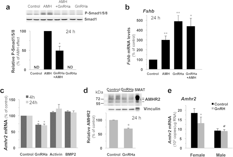
Anti-Müllerian hormone (AMH) contributes to male sexual differentiation and acts on gonads of both sexes. Identification of AMH receptivity in both pituitary and brain has led to the intriguing idea that AMH participates to the hypothalamic-pituitary control of reproduction, however in vivo experimental evidence is still lacking. We show that AMH stimulates secretion and pituitary gene expression of the gonadotropin FSH in vivo in rats. AMH action is sex-dependent, being restricted to females and occurring before puberty. Accordingly, we report higher levels of pituitary AMH receptor transcripts in immature females. We show that AMH is functionally coupled to the Smad pathway in LβT2 gonadotrope cells and dose-dependently increases Fshb transcript levels. Furthermore, AMH was shown to establish complex interrelations with canonical FSH regulators as it cooperates with activin to induce Fshb expression whereas it reduces BMP2 action. We report that GnRH interferes with AMH by decreasing AMH receptivity in vivo in females. Moreover, AMH specifically regulates FSH and not LH, indicating that AMH is a factor contributing to the differential regulation of gonadotropins. Overall, our study uncovers a new role for AMH in regulating gonadotrope function and suggests that AMH participates in the postnatal elevation of FSH secretion in females.
Figures


Similar articles
-
Cell-specific transcriptional regulation of follicle-stimulating hormone-beta by activin and gonadotropin-releasing hormone in the LbetaT2 pituitary gonadotrope cell model.Endocrinology. 2001 Jun;142(6):2284-95. doi: 10.1210/endo.142.6.8185. Endocrinology. 2001. PMID: 11356674
-
Anti-Müllerian hormone receptor type 2 is expressed in gonadotrophs of postpubertal heifers to control gonadotrophin secretion.Reprod Fertil Dev. 2018 Aug;30(9):1192-1203. doi: 10.1071/RD17377. Reprod Fertil Dev. 2018. PMID: 29533759
-
Discovery of new receptors regulating luteinizing hormone and follicle-stimulating hormone secretion by bovine gonadotrophs to explore a new paradigm for mechanisms regulating reproduction.J Reprod Dev. 2020 Aug 20;66(4):291-297. doi: 10.1262/jrd.2020-012. Epub 2020 Apr 6. J Reprod Dev. 2020. PMID: 32249236 Free PMC article. Review.
-
Anti-Müllerian hormone and its receptor are detected in most gonadotropin-releasing-hormone cell bodies and fibers in heifer brains.Domest Anim Endocrinol. 2020 Jul;72:106432. doi: 10.1016/j.domaniend.2019.106432. Epub 2020 Jan 3. Domest Anim Endocrinol. 2020. PMID: 32169754
-
Pituitary gonadotroph-specific patterns of gene expression and hormone secretion.Curr Opin Pharmacol. 2022 Oct;66:102274. doi: 10.1016/j.coph.2022.102274. Epub 2022 Aug 19. Curr Opin Pharmacol. 2022. PMID: 35994915 Free PMC article. Review.
Cited by
-
Serum Concentrations of AMH and E2 and Ovarian and Uterine Traits in Gilts.Animals (Basel). 2019 Oct 15;9(10):811. doi: 10.3390/ani9100811. Animals (Basel). 2019. PMID: 31619004 Free PMC article.
-
Anti-Mullerian Hormone Induces Foxo1 and Sirt1 Genes Expression in Mouse Ovary.Curr Pharm Biotechnol. 2025;26(6):872-877. doi: 10.2174/0113892010293250240917143811. Curr Pharm Biotechnol. 2025. PMID: 39328137
-
Endocrine Responses to Triptorelin in Healthy Women, Women With Polycystic Ovary Syndrome, and Women With Hypothalamic Amenorrhea.J Clin Endocrinol Metab. 2023 Jun 16;108(7):1666-1675. doi: 10.1210/clinem/dgad026. J Clin Endocrinol Metab. 2023. PMID: 36653328 Free PMC article. Clinical Trial.
-
A screen of repurposed drugs identifies AMHR2/MISR2 agonists as potential contraceptives.Proc Natl Acad Sci U S A. 2022 Apr 12;119(15):e2122512119. doi: 10.1073/pnas.2122512119. Epub 2022 Apr 5. Proc Natl Acad Sci U S A. 2022. PMID: 35380904 Free PMC article.
-
Regulation of the neuroendocrine axis in male rats by soy-based diets is independent of age and due specifically to isoflavone action†.Biol Reprod. 2020 Oct 5;103(4):892-906. doi: 10.1093/biolre/ioaa101. Biol Reprod. 2020. PMID: 32520353 Free PMC article.
References
Publication types
MeSH terms
Substances
LinkOut - more resources
Full Text Sources
Other Literature Sources
Molecular Biology Databases
