. 2016 Mar 30;8(332):332ra42.
doi: 10.1126/scitranslmed.aaf1164.

Somatic PIK3CA mutations as a driver of sporadic venous malformations

Affiliations

Somatic PIK3CA mutations as a driver of sporadic venous malformations

Pau Castel et al. Sci Transl Med. .

Abstract

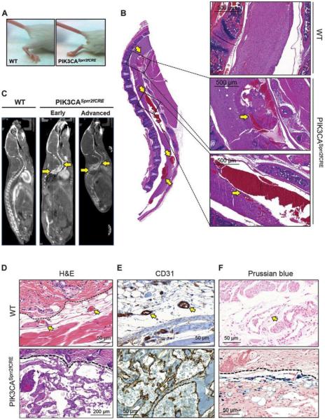
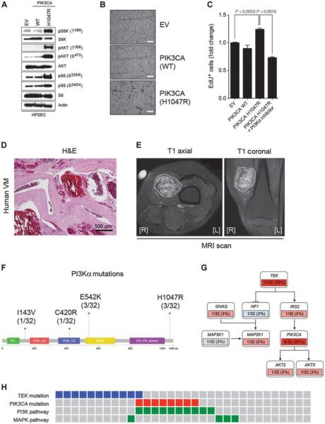
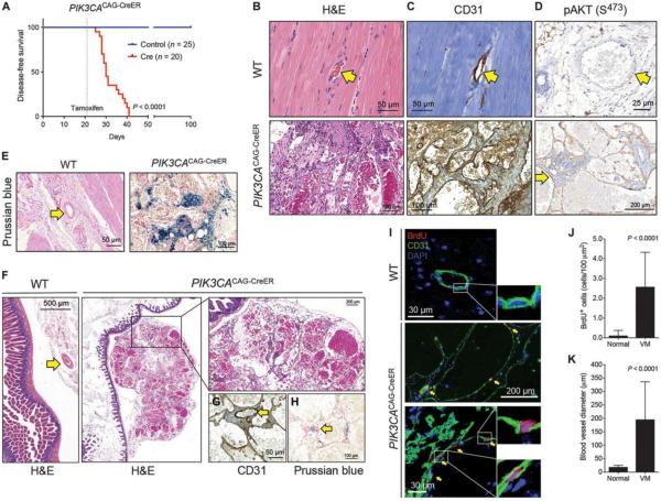
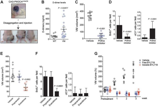
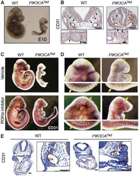
Venous malformations (VM) are vascular malformations characterized by enlarged and distorted blood vessel channels. VM grow over time and cause substantial morbidity because of disfigurement, bleeding, and pain, representing a clinical challenge in the absence of effective treatments (Nguyenet al, 2014; Uebelhoeret al, 2012). Somatic mutations may act as drivers of these lesions, as suggested by the identification of TEK mutations in a proportion of VM (Limayeet al, 2009). We report that activating PIK3CA mutations gives rise to sporadic VM in mice, which closely resemble the histology of the human disease. Furthermore, we identified mutations in PIK3CA and related genes of the PI3K (phosphatidylinositol 3-kinase)/AKT pathway in about 30% of human VM that lack TEK alterations. PIK3CA mutations promote downstream signaling and proliferation in endothelial cells and impair normal vasculogenesis in embryonic development. We successfully treated VM in mouse models using pharmacological inhibitors of PI3Kα administered either systemically or topically. This study elucidates the etiology of a proportion of VM and proposes a therapeutic approach for this disease.


Figures

Fig. 1
Fig. 1. PIK3CASprr2fCRE mice develop spinal vascular malformations
(A) Hindlimb paresis phenotype observed in PIK3CASprr2fCRE mice. WT, wild type. (B) Gross and detailed histology of the spinal cord of PIK3CASprr2fCRE mice compared to a normal WT spine. Arrows indicate the multiple focal hemorrhages found in the spinal cord. (C) microCT (micro-computed tomography) scan of a WT mouse compared to PIK3CASprr2fCRE mice littermates showing an early and an advanced phenotype. Arrows indicate the slow flow and extravasation lesions observed in the spinal cord. (D) Hematoxylin and eosin (H&E) histology from normal skin and cutaneous VM. Dashed line delimits the dermis (lower) from the epidermis (upper). Arrows indicate normal blood vessels. (E) CD31 immunohistochemistry (IHC) of the skin VM lesions. Arrows indicate normal blood vessels. (F) Prussian blue staining. Dashed line delimits the dermis (lower) from the epidermis (upper). Arrow indicates normal blood vessel.
Fig. 2
Fig. 2. PIK3CA mutations cause sporadic VM in humans
(A) Western blot of human skin ECs infected with empty vector (EV), PIK3CA WT, and H1047R mutation probed with the indicated antibodies. Cells were serum-starved overnight before lysis. S6K, S6 kinase; pS6K, phosphorylated S6K; pS6, phosphorylated S6; pAKT, phospshorylated AKT; HFSEC, human female skin endothelial cell. (B) Representative images from the tube formation assays of primary ECs infected with EV, PIK3CA WT, and H1047R mutation and serum-starved overnight before seeding. Pictures were taken 6 hours after seeding. Note the reticular network formed in the EV and PIK3CA WT cells that fails to form in the PIK3CA H1047R mutant cells. Scale bars, 200 μm. (C) EdU incorporation assay quantification of ECs infected with EV, PIK3CA WT, and H1047R mutation, serum-starved overnight, and labeled with EdU for 4 hours. Graph shows mean fold change ± SEM. n = 3 biological replicates. P values were calculated using Student’s t test. (D) H&E staining highlighting the representative morphology of one of the human VM patients sequenced in this study. Blood pools and thick mural cell layer are evident in the histological sections of these patients. (E) Characteristic MRI scan from an intramuscular sporadic VM patient sequenced in this study. T1 axial and coronal sections are shown. Dashed line delimits the radiological extension of the malformation. [R] and [L] indicate right and left, respectively. (F) PI3Kα domains and specific sites found to be mutated in this study. The p85-binding domain is represented in green, the Rasbinding domain in red, the C2 domain in blue, the helical domain in yellow, and the kinase domain in purple. aa, amino acid. (G) Schematic pathway depicting TIE2, PI3K, and MAPK pathway gene components found to be mutated in sporadic VM by MSK-IMPACT in this study. Activating mutations are indicated in red and inactivating mutations in blue. Dark red designates mutations found in more than 20% of the patients. Unknown mutations are shown in gray. (H) Mutual exclusivity of the gene mutations present in the TEK and PIK3CA pathways. The activating mutations in TEK are indicated in blue, and the activating mutations in PIK3CA are in red. The alterations affecting genes involved in the PI3K or MAPK pathway are represented in green.
Fig. 3
Fig. 3. Ubiquitous expression of PIK3CAH1047R induces VM
(A) Disease-free survival plot of PIK3CACAG-CreER (n = 20) and PIK3CAWT (n = 25) littermates on tamoxifen diet assessed by the appearance of visible cutaneous VM. Dotted line represents the time when tamoxifen diet was administered (day 21). P value was calculated using log-rank test. (B) H&E staining of a representative normal blood vessel and a VM lesion developed in the skin of PIK3CACAG-CreER mice. Arrow indicates normal blood vessel. (C) CD31 IHC staining showing positivity for the ECs of a normal blood vessel and VM. Note that erythrocytes exhibit nonspecific staining. Arrow indicates normal blood vessel. (D) Phosphorylated AKT (S473) IHC. The arrow in the bottom panel indicates lining ECs that show positivity for the staining. Note the negativity for phosphorylated AKT in the normal blood vessel (top, indicated by an arrow). (E) Prussian blue staining of normal blood vessels and the PIK3CACAG-CreER mouse skin VM lesions. Arrow indicates a normal blood vessel. (F) Histological representation of mesenteric vasculature and VM harvested during necropsy and detailed view to highlight the blood pools observed in the preparations. Arrows indicate normal blood vessels. (G and H) CD31 (G) and Prussian blue (H) positivity for the VM described in (F). (I) BrdU incorporation (red) in PIK3CACAG-CreER VM compared to normal blood vessels. CD31 (green) and DAPI (4′,6-diamidino-2-phenylindole) (blue) show ECs and nuclei, respectively. Note the encased BrdU-positive nuclei in the CD31-positive lining EC layer. (J) Quantification of BrdU-positive nuclei in normal blood vessels and VM. P value was calculated using Student’s t test. Graph shows means ± SD. n = 45 fields from five biological replicates. (K) Morphological quantification of the maximal blood vessel diameter of normal vessels and VM. P value was calculated using Student’s t test. Graph shows means ± SD. n = 45 fields from five biological replicates.
Fig. 4
Fig. 4. PI3K inhibitors are effective for the treatment of VM
(A) Schematic representation and images from the allotransplantation assays. (B) Quantification of plasma D-dimers measured in animals with or without VM. P value was calculated using Student’s t test. (C) VM volume measured in PIK3CACAG-CreER VM-derived allografts treated with vehicle or PI3Kα inhibitor for 1 week [BYL719, 50 mg kg−1, daily, per os (po)]. P value was calculated using Student’s t test. (D) Quantification of BrdU incorporation and cleaved caspase-3 (clC3) in CD31-positive cells from (B). P value was calculated using Student’s t test. n = 10. (E) VM volume measured in PIK3CACAG-CreER VM-derived allografts treated with vehicle, everolimus (10 mg kg−1, daily, po), or propranolol (40 mg kg−1, daily, po) for 1 week. P value was calculated using Student’s t test. (F) Quantification of BrdU incorporation and cleaved caspase-3 in CD31-positive cells from (E). P value was calculated using Student’s t test. n = 8. (G) VM volume in PIK3CACAG-CreER VM-derived allografts treated topically with BYL719 at 1% (w/w) using two different formulations (free and soluble BYL719) for 3 weeks. The pretreatment time point indicates when the treatment was started. All treatments in weeks 1, 2, and 3 have a P < 0.001 as compared to the vehicle control VM. P values were calculated using Student’s t test.
Fig. 5
Fig. 5. PIK3CAH1047R impairs embryonic angiogenesis
(A) Embryonic phenotype observed in PIK3CAWT (left) and PIK3CATie2-Cre (right) littermates at E10. For morphological studies, a minimum of four dissections for each genotype were performed yielding n ≥ 15 embryos. (B) CD31 staining of coronal sections from PIK3CAWT and PIK3CATie2-Cre embryos at E9.5. Arrowheads indicate blood vessel enlargement defects in the meningeal vessels (upper panel), and arrows indicate the defects in the intersomitic vessels (lower panel). For CD31 histologic studies, a minimum of four embryos for each phenotype obtained from two different dissections were used. Scale bars, 100 μm. (C) Whole-embryo CD31 staining of PIK3CAWT and PIK3CATie2-Cre embryos from mice treated with vehicle or PI3Kα inhibitor (BYL719, 50 mg kg−1, daily, po; 48, 24, and 2 hours before embryos are harvested). For CD31 histologic studies, a minimum of four embryos for each condition were used. (D) Detailed view of the cephalic and intersomitic blood vessels from (C). Arrow indicates defects in the meningeal (upper panel) and the intersomitic vessels (lower panel). (E) CD31 coronal sections from embryos in (D). CV, cardinal vein; DA, dorsal aorta. Scale bars, 100 μm.

Similar articles

Cited by

References

    1. Brouillard P, Vikkula M. Genetic causes of vascular malformations. Hum. Mol. Gen. 2007;16:R140–R149. - PubMed
    1. Brouillard P, Vikkula M. Vascular malformations: Localized defects in vascular morphogenesis. Clin. Genet. 2003;63:340–351. - PubMed
    1. Dompmartin A, Vikkula M, Boon LM. Venous malformation: Update on aetiopathogenesis, diagnosis and management. Phlebology. 2010;25:224–235. - PMC - PubMed
    1. Nguyen H-L, Boon LM, Vikkula M. Genetics of vascular malformations. Semin. Pediatr. Surg. 2014;23:221–226. - PubMed
    1. Uebelhoer M, Boon LM, Vikkula M. Vascular anomalies: From genetics toward models for therapeutic trials. Cold Spring Harb. Perspect. Med. 2012;2:a009688. - PMC - PubMed

Publication types

MeSH terms