. 2016 Mar 30;8(332):332ra43.
doi: 10.1126/scitranslmed.aad9982.

Somatic activating mutations in Pik3ca cause sporadic venous malformations in mice and humans

Affiliations

Somatic activating mutations in Pik3ca cause sporadic venous malformations in mice and humans

Sandra D Castillo et al. Sci Transl Med. .

Abstract

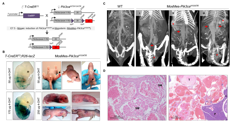
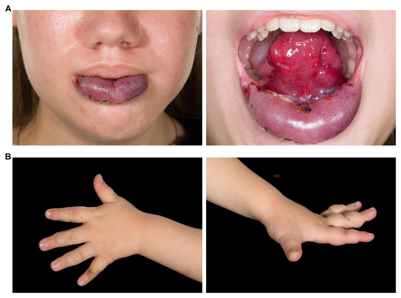
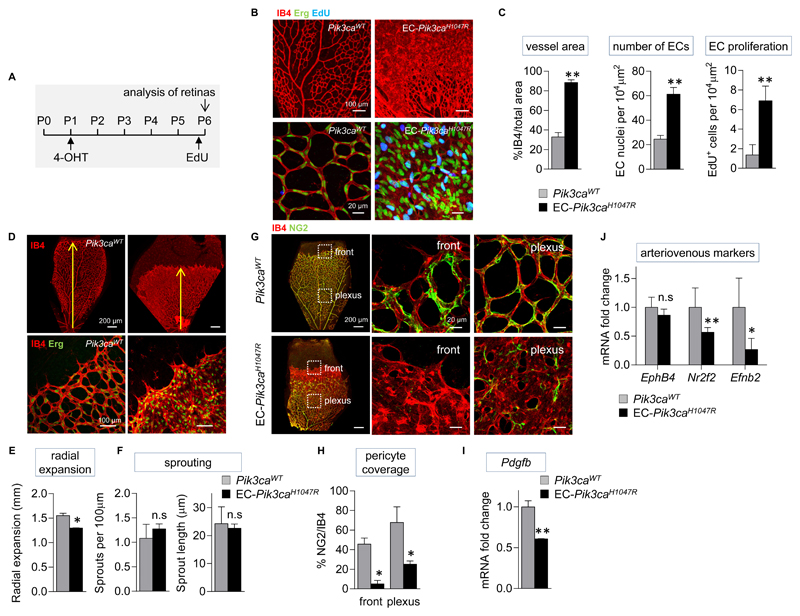
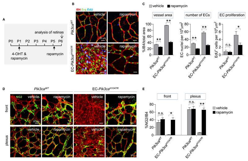
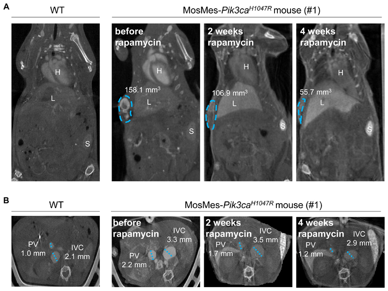
Venous malformations (VMs) are painful and deforming vascular lesions composed of dilated vascular channels, which are present from birth. Mutations in the TEK gene, encoding the tyrosine kinase receptor TIE2, are found in about half of sporadic (nonfamilial) VMs, and the causes of the remaining cases are unknown. Sclerotherapy, widely accepted as first-line treatment, is not fully efficient, and targeted therapy for this disease remains underexplored. We have generated a mouse model that faithfully mirrors human VM through mosaic expression of Pik3ca(H1047R), a constitutively active mutant of the p110α isoform of phosphatidylinositol 3-kinase (PI3K), in the embryonic mesoderm. Endothelial expression of Pik3ca(H1047R)resulted in endothelial cell (EC) hyperproliferation, reduction in pericyte coverage of blood vessels, and decreased expression of arteriovenous specification markers. PI3K pathway inhibition with rapamycin normalized EC hyperproliferation and pericyte coverage in postnatal retinas and stimulated VM regression in vivo. In line with the mouse data, we also report the presence of activating PIK3CA mutations in human VMs, mutually exclusive with TEK mutations. Our data demonstrate a causal relationship between activating Pik3ca mutations and the genesis of VMs, provide a genetic model that faithfully mirrors the normal etiology and development of this human disease, and establish the basis for the use of PI3K-targeted therapies in VMs.


Conflict of interest statement

Competing financial interests

B.V. is consultant to Karus Therapeutics (Oxford, UK). M.F.L. serves as a board member of the International Society for Magnetic Resonance in Medicine (British Chapter). All other authors declare that they have no competing interests.

Figures

Figure 1
Figure 1. Mosaic expression of Pik3caH1047R in embryonic mouse mesoderm induces vascular malformations.
(A) Genetic strategy for mosaic Pik3caH1047R induction in the embryonic mesoderm. T-CreERT2 mice were crossed with Pik3caWT/H1047R mice that have a germline Pik3ca allele with a conditional H1047R mutation in exon 20. Mosaic recombination in the mesoderm was induced by a single intra-peritoneal injection of a low dose of 4-OHT to pregnant mice at E7.5. (B) Left, E12.5 T-CreERT2;R26-lacZ mouse embryos from pregnant mice injected with the indicated dose of 4-OHT at E7.5 and stained for β-galactosidase (β-gal) activity. Right, Representative photographs of P1-P5 MosMes-Pik3caH1047R pups with congenital vascular malformations (indicated by arrows in the top panel), born to pregnant mice injected with the indicated 4-OHT dose at E7.5. (C) CT-A scans of adult mice four hours after intravenous injection of gold nanoparticles. The WT mouse (left) shows normal vascular anatomy, whereas MosMes-Pik3caH1047R mice display dilatation of the left common iliac vein (arrow) and VM in the urogenital area (asterisk) (mouse #1); subcutaneous VM (asterisk) and dilatation of the inferior vena cava (arrow) (mouse #2); subcutaneous and urogenital VMs (asterisks) and dilatation of the inferior vena cava and portal vein (arrows) (mouse #3). (D) Representative hematoxylin and eosin (H&E)-stained sections of subcutaneous (left) and deep mesenteric (right) VMs in MosMes-Pik3caH1047R mice, showing abnormal, enlarged, and irregular vascular channels, most containing blood and organising fibrin thrombi (T), some interposing between skeletal muscle (SM) and other tissue structures. No cytological atypia was observed. (P, pancreas).
Figure 2
Figure 2. PIK3CA is mutated in human VMs.
(A) Patient 9 showing VMs (PIK3CAH1047L) in the lower lip and tongue. (B) Patient 13 with VM (PIK3CAE545K) in the left hand.
Figure 3
Figure 3. Endothelial activation of Pik3ca promotes hyperproliferation in ECs and impairs pericyte coverage.
(A) Schematic of the 4-OHT and EdU administration regime used. (B) Representative flat-mounted Pik3caWT and EC-Pik3caH1047R P6 retinas stained with IB4 (red, revealing ECs) and antibody to the Erg transcription factor (nuclear marker of ECs; green) and labelled with EdU (blue). (C) Quantitative analysis of the retina vessel area (assessed by IB4 staining), EC numbers (assessed by staining for Erg), and number of proliferating ECs (cells positive for both EdU and Erg). Data represent mean ± SEM. **p ≤ 0.01 (Mann-Whitney U test). n=6/genotype. (D) Representative flat-mounted control and EC-Pik3caH1047R P6 retinas stained with IB4 and antibody to the Erg transcription factor. (E) Quantification of the radial expansion of vasculature in retinas. Data represent mean ± SEM. *p < 0.05 (Mann-Whitney U test). n=6/genotype. (F) Quantification of the number of sprouts at the vascular front per unit length, and the length of sprouts. Data represent mean ± SEM. n.s., not significant, p > 0.05 (Mann-Whitney U test). n=6/genotype. (G) Flat-mounted Pik3caWT and EC-Pik3caH1047R retinas showing vasculature (IB4; red) and pericytes (stained for NG2, a membrane proteoglycan found in pericytes; green). Right, higher magnification of highlighted sections. (H) Quantification of pericyte coverage in the vasculature of retinas (assessed by % of NG2 staining relative to IB4 staining). Data represent mean ± SEM. *p < 0.05 (Mann-Whitney U test). n=6/genotype. (I) Pdgfb mRNA expression in EC-Pik3caH1047R P6 retinas. Data represent mean ± SEM. **p < 0.01 (Mann-Whitney U test). n=5/genotype. (J) Ephb4, Nr2f2, and Efnb2 mRNA expression in EC-Pik3caH1047R P6 retinas. Data represent mean ± SEM. n.s., not significant, p > 0.05, *p < 0.05, **p < 0.01 (Mann-Whitney U test). n=5/genotype.
Figure 4
Figure 4. Rapamycin reduces Pik3caH1047R EC hyperproliferation and prevents loss of pericyte coverage in postnatal retinas.
(A) Schematic of the 4-OHT and rapamycin administration regime used for analysis of retinal angiogenesis. (B) Representative flat-mounted P6 retinas from vehicle and rapamycin-treated Pik3caWT and EC-Pik3caH1047R pups. Retinas are stained with IB4 (red) and antibody to the Erg transcription factor (green) and labeled with EdU (blue). (C) Quantitative analysis of the retinal vessel area (assessed by IB4 staining), EC numbers (assessed by staining for Erg), and number of proliferating ECs (cells positive for both EdU and Erg). Data represent mean ± SEM. n.s., not significant, p>0.05, *p < 0.05, **p < 0.01 (Mann-Whitney U test). n=6/genotype. (D) Representative flat-mounted P6 retinas from untreated Pik3caWT and EC-Pik3caH1047R pups and rapamycin-treated EC-Pik3caH1047R pups showing vasculature (IB4; red) and pericytes (stained for NG2; green). (E) Quantitative analysis of pericyte coverage in the vascular front and plexus of retinas (assessed by % of NG2 staining relative to IB4 staining). Data represent mean ± SEM. n.s., not significant, p > 0.05, *p < 0.05, **p < 0.01 (Mann-Whitney U test). n=6/genotype.
Figure 5
Figure 5. Rapamycin induces regression of Pik3caH1047R-driven VMs in vivo.
(A) Measurement of the volume of the subcutaneous VM (circled in blue) from CT-A images of mouse #1 (Table 2), showing the volume of the VM. H, heart; L, liver; S, spleen. (B) Measurement of the average diameter of the inferior vena cava and portal vein from CT-A images of an untreated WT mouse and a MosMes-Pik3caH1047R littermate mouse, before and after 2 and 4 weeks of rapamycin treatment. IVC, inferior vena cava; PV, portal vein.

References

    1. Vanhaesebroeck B, Stephens L, Hawkins P. PI3K signalling: the path to discovery and understanding. Nature reviews. Molecular cell biology. 2012;13:195–203. - PubMed
    1. Samuels Y, et al. High frequency of mutations of the PIK3CA gene in human cancers. Science. 2004;304:554. - PubMed
    1. Kurek KC, et al. Somatic mosaic activating mutations in PIK3CA cause CLOVES syndrome. American journal of human genetics. 2012;90:1108–1115. - PMC - PubMed
    1. Lindhurst MJ, et al. Mosaic overgrowth with fibroadipose hyperplasia is caused by somatic activating mutations in PIK3CA. Nature genetics. 2012;44:928–933. - PMC - PubMed
    1. Riviere JB, et al. De novo germline and postzygotic mutations in AKT3, PIK3R2 and PIK3CA cause a spectrum of related megalencephaly syndromes. Nature genetics. 2012;44:934–940. - PMC - PubMed

Publication types

MeSH terms