. 2016 Mar 10:12:1744806916637699.
doi: 10.1177/1744806916637699. Print 2016.

Involvement of NIPSNAP1, a neuropeptide nocistatin-interacting protein, in inflammatory pain

Affiliations

Involvement of NIPSNAP1, a neuropeptide nocistatin-interacting protein, in inflammatory pain

Kazuya Okamoto et al. Mol Pain. .

Abstract

Background: Chronic pain associated with inflammation is an important clinical problem, and the underlying mechanisms remain poorly understood. 4-Nitrophenylphosphatase domain and nonneuronal SNAP25-like protein homolog (NIPSNAP) 1, an interacting protein with neuropeptide nocistatin, is implicated in the inhibition of tactile pain allodynia. Although nocistatin inhibits some inflammatory pain responses, whether NIPSNAP1 affects inflammatory pain appears to be unclear. Here, we examined the nociceptive behavioral response of NIPSNAP1-deficient mice and the expression of NIPSNAP1 following peripheral inflammation to determine the contribution of NIPSNAP1 to inflammatory pain.

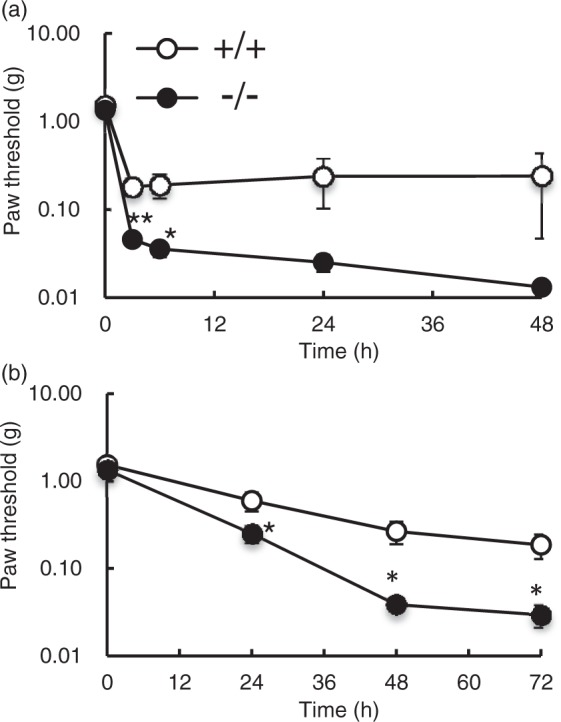
Results: Nociceptive behavioral response increased in phase II of the formalin test, particularly during the later stage (26-50 min) in NIPSNAP1-deficient mice, although the response during phase I (0-15 min) was not significantly different between the deficient and wild-type mice. Moreover, phosphorylation of extracellular signal-related kinase was enhanced in the spinal dorsal horn of the deficient mice. The prolonged inflammatory pain induced by carrageenan and complete Freund's adjuvant was exacerbated in NIPSNAP1-deficient mice. NIPSNAP1 mRNA was expressed in small- and medium-sized neurons of the dorsal root ganglion and motor neurons of the spinal cord. In the formalin test, NIPSNAP1 mRNA was slightly increased in dorsal root ganglion but not in the spinal cord. In contrast, NIPSNAP1 mRNA levels in dorsal root ganglion were significantly decreased during 24-48 h after carrageenan injection. Prostaglandin E2, a major mediator of inflammation, stimulated NIPSNAP1 mRNA expression via the cAMP-protein kinase A signaling pathway in isolated dorsal root ganglion cells.

Conclusions: These results suggest that changes in NIPSNAP1 expression may contribute to the pathogenesis of inflammatory pain.

Keywords: NIPSNAP1; gene expression; inflammatory pain; prostaglandin E2.


Figures

Figure 1.
Figure 1.
Effects of NIPSNAP1 deficiency on pain sensitivity. Response latencies of wild-type (+/+) and NIPSNAP1−/− (−/−) mice on the hot plate at 50℃ and 55℃ (a) and their response to a radiant heat stimulus (b). Data are expressed as the mean ± SEM (n = 9–12). The paw threshold of wild-type and NIPSNAP1−/− mice by the first response method (c) and the 50% withdrawal threshold by the up-down method (d) induced by mechanical stimuli using the von Frey filaments (n = 7). Heat stimulus (55℃, 1 min) induced ERK phosphorylation (red) in the spinal dorsal horn of wild-type and NIPSNAP1−/− mice. Representative results (e) and number of p-ERK-positive neurons (f) in the spinal dorsal horn at 5 min after heat stimulation. p-ERK-positive neurons were quantified in four slices prepared from the three separated mice (mean ± SEM).
Figure 2.
Figure 2.
Pain sensitization induced in NIPSNAP1−/− mice by peripheral injection of formalin. Time spent licking and biting per 5 min is plotted versus time after peripheral injection of 2% formalin into the right hindpaw of wild-type and NIPSNAP1−/− mice. Data are expressed as the mean ± SEM (n = 11–13). **p < 0.01, *p < 0.05, versus wild-type value. (b) Cumulative time spent licking and biting during phase I (0–15 min), phase II-e (16–25 min), and phase II-l (26–50 min). *p < 0.05, versus wild-type value. (c) Formalin injected into the hind paw of wild-type and NIPSNAP1−/− mice induced ERK phosphorylation (red) in the ipsilateral dorsal horn of the spinal cord. Representative immunohistochemical data at the times indicated after formalin injection. (d) Number of p-ERK-positive neurons in the dorsal horn of spinal cord induced by formalin. p-ERK-positive neurons were quantified in four to eight slices prepared from the three to six separated mice (mean ± SEM). **p < 0.01, *p < 0.05, versus wild-type value.
Figure 3.
Figure 3.
Effect of NIPSNAP1 deficiency on the carrageenan-induced inflammatory responses in the paws. In the paws at 5 h after the peripheral carrageenan injection of wild-type and NIPSNAP1−/− mice, the diameter (a), the level of MCP-1 (b), and the numbers of F4/80-positive macrophages, and Gr-1-positive neutrophils (c, d) were measured. Representative results (c) and number of F4/80 - and Gr-1-positive cells were quantified in two slices prepared from the three separated mice (d). Data are expressed as the mean ± SEM (a, n = 8; b, n = 4; c, n = 6).
Figure 4
Figure 4
Pain sensitization induced by peripheral injection of NIPSNAP1−/− mice with carrageenan and CFA. Paw withdrawal latencies of mice injected with 1% carrageenan (a) or CFA (b) using von Frey filaments. Data are expressed as the mean ± SEM (n = 7). **p < 0.01, *p < 0.05, versus wild-type value.
Figure 5.
Figure 5.
Distribution of NIPSNAP1 mRNA. (a) RT-PCR analysis of NIPSNAP1 mRNA expression in the spinal cord, paw, and DRG. GAPDH served as the control. (b) In situ hybridization analysis of NIPSNAP1 mRNA expression in the spinal cords and dorsal root ganglia of wild-type and NIPSNAP1−/− mice.
Figure 6.
Figure 6.
Analysis of NIPSNAP1 mRNA levels following the induction of inflammation. Levels of NIPSNAP1 mRNA in DRG and spinal cord induced by injection into the dorsal surface of hind paw of 2% formalin (a) or 1% carrageenan (n). The upper and lower panels show representative results of RT-PCR and real-time PCR analysis, respectively. Data are expressed as the mean ± SEM (n = 3). **p < 0.01, *p < 0.05, injected versus controls.
Figure 7
Figure 7
Analysis of NIPSNAP1 mRNA expression induced by PGE2. (a) RT-PCR and real-time PCR analysis of NIPSNAP1 expression induced by 5 µM PGE2 in DRG cells. (b) RT-PCR analysis of the expression of mRNA encoding EP subtypes in DRG cells. White arrowhead shows EP3β. (c) Effect of EP agonists on the expression of NIPSNAP1 mRNA. DRG cells were incubated for 1 h in the presence of each EP agonist (1 µM), and subjected to RT-PCR and real-time PCR analysis. (d) Effect of the protein kinase A inhibitor H-89 on the PGE2-induced expression of NIPSNAP1 mRNA. DRG cells incubated with 5 µM PGE2 for 1 h in the absence and presence of 20 µM H-89 were subjected to RT-PCR and real-time PCR analysis. The upper and lower panels show representative results of RT-PCR and real-time PCR analysis, respectively. Data are expressed as the mean ± SEM (n = 3–4). **p < 0.01, *p < 0.05, treated versus untreated cells. ##p < 0.01, treated versus PGE2-treated cells.
Figure 8.
Figure 8.
Effect of cyclooxygenase inhibitor on the formalin-induced NIPSNAP1 mRNA increase in DRG. Aspirin (300 µg/g) was administered orally to mice for 1 h before the peripheral formalin injection. Data are expressed as the mean ± SEM (n = 3–6). *p < 0.05, versus vehicle-administered mice.

References

    1. Tummala H, Li X, Homayouni R. Interaction of a novel mitochondrial protein, 4-nitrophenylphosphatase domain and non-neuronal SNAP25-like protein homolog 1 (NIPSNAP1), with the amyloid precursor protein family. Eur J Neurosci 2010; 31: 1926–1934. - PubMed
    1. Nautiyal M, Sweatt AJ, MacKenzie JA, et al. Neuronal localization of the mitochondrial protein NIPSNAP1 in rat nervous system. Eur J Neurosci 2010; 32: 560–569. - PubMed
    1. Okuda-Ashitaka E, Minami T, Tsubouchi S, et al. Identification of NIPSNAP1 as a nocistatin-interacting protein involving pain transmission. J Biol Chem 2012; 287: 10403–10413. - PMC - PubMed
    1. Satoh K, Takeuchi M, Oda Y, et al. Identification of activity-regulated proteins in the postsynaptic density fraction. Genes Cells 2002; 7: 187–197. - PubMed
    1. Takata A, Kakiuchi C, Ishiwata M, et al. Behavioral and gene expression analyses in heterozygous XBP1 knockout mice: possible contribution of chromosome 11qA1 locus to prepulse inhibition. Neurosci Res 2010; 68: 250–255. - PubMed

Publication types

MeSH terms

LinkOut - more resources