Editorial
. 2016 Jun 1;115(6):3140-5.
doi: 10.1152/jn.00089.2016. Epub 2016 Mar 30.

The functional organization of human epileptic hippocampus

Affiliations
Editorial

The functional organization of human epileptic hippocampus

Petr Klimes et al. J Neurophysiol. .

Abstract

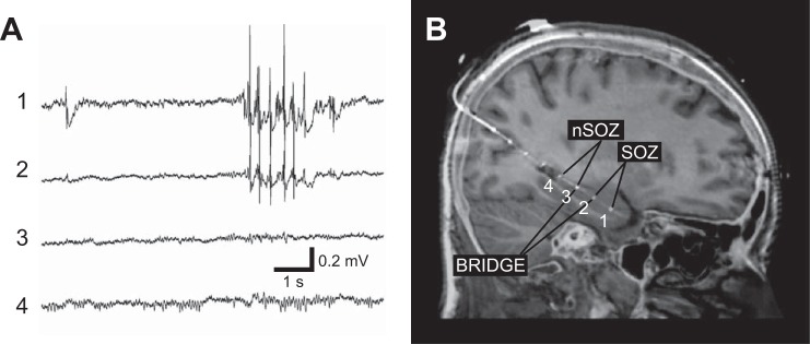
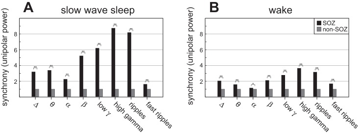
The function and connectivity of human brain is disrupted in epilepsy. We previously reported that the region of epileptic brain generating focal seizures, i.e., the seizure onset zone (SOZ), is functionally isolated from surrounding brain regions in focal neocortical epilepsy. The modulatory effect of behavioral state on the spatial and spectral scales over which the reduced functional connectivity occurs, however, is unclear. Here we use simultaneous sleep staging from scalp EEG with intracranial EEG recordings from medial temporal lobe to investigate how behavioral state modulates the spatial and spectral scales of local field potential synchrony in focal epileptic hippocampus. The local field spectral power and linear correlation between adjacent electrodes provide measures of neuronal population synchrony at different spatial scales, ∼1 and 10 mm, respectively. Our results show increased connectivity inside the SOZ and low connectivity between electrodes in SOZ and outside the SOZ. During slow-wave sleep, we observed decreased connectivity for ripple and fast ripple frequency bands within the SOZ at the 10 mm spatial scale, while the local synchrony remained high at the 1 mm spatial scale. Further study of these phenomena may prove useful for SOZ localization and help understand seizure generation, and the functional deficits seen in epileptic eloquent cortex.

Keywords: behavioral state; connectivity; epilepsy; intracranial EEG; seizure onset zone.


Figures

Fig. 1.
Fig. 1.
Intracranial EEG and MRI-CT coregistered electrodes. A: the intracranial recording, i.e., local field potentials (LFP), from an 8 contact depth electrode implanted along the long axis of the hippocampus via a posterior approach. The anterior most contact 1 targeting the anterior hippocampus and contact 2 show interictal epileptiform spikes. In this patient contacts 1–4 span the length of the hippocampus. B: the seizure onset zone (SOZ) was determined by recording spontaneous seizures and contacts outside the SOZ are denoted as non-SOZ (nSOZ). The contacts that bridge the SOZ and non-SOZ (here 3 and 2) are labeled as bridge pairs.
Fig. 2.
Fig. 2.
Ten-millimeter spatial scale synchrony (correlation, bipolar power), and behavioral state modulation (top). The linear correlation between electrode pairs located in SOZ, non-SOZ, and bridging areas in the delta (1–4 Hz), theta (4–8 Hz), alpha (8–12 Hz), beta (12–20 Hz), low gamma (20–55 Hz), high gamma (65–80 Hz), ripples (80–250 Hz), and fast ripples (250–600 Hz) frequency bands. During sleep (left top, A), the correlation is increased in SOZ compared with non-SOZ at frequencies delta-beta, but exhibits a cross-over to low connectivity in higher frequency bands within the SOZ. The connectivity between electrodes that bridge SOZ and non-SOZ (bridging electrode pairs) exhibit lower connectivity compared with SOZ and non-SOZ, except at ripples and fast ripples. Similarly, during wakefulness (right top, B) the connectivity between bridging electrodes is reduced compared with electrodes within the SOZ and non-SOZ (bottom) The bipolar power is calculated from the difference of two adjacent electrode LFPs. Relative changes of bipolar power between groups − bipolar power values at each frequency are normalized to power outside SOZ = 1. During slow-wave sleep (left bottom, C) the bipolar power is increased in SOZ compared with non-SOZ for all frequency bands, supporting a relative statistical independence of the activity at adjacent electrodes in the SOZ compared with electrodes in the non-SOZ. The highest power is in high gamma. The bipolar power in areas bridging SOZ and non-SOZ is increased compared with non-SOZ areas for all frequency bands except fast ripples, supporting that the connectivity between SOZ and non-SOZ is reduced. During wake (right bottom, D) the bipolar power is the highest in SOZ in high gamma. The bipolar power in SOZ and bridging areas is lower than non-SOZ in theta and alpha band. The differences between bipolar power in SOZ-non-SOZ and bridges-non-SOZ are lower for all frequency bands compared with slow-wave sleep. Statistical difference between groups is observed at two levels, *P < 0.05 and **P < 0.01.
Fig. 3.
Fig. 3.
One-millimeter spatial scale synchrony (unipolar power), and behavioral state modulation. The LFP unipolar spectral power from a single electrode can be interpreted as a local (∼1 mm) measure of neuronal synchrony (power normalized to non-SOZ = 1). The LFP unipolar power is increased in SOZ compared with non-SOZ for all frequency bands, during both behavioral states [slow-wave sleep (A), wake (B)]. The difference between SOZ and non-SOZ is reduced during wakefulness in alpha band. In general, differences between SOZ and non-SOZ were smaller during wake state. Statistical difference between groups is observed at two levels, *P < 0.05 and **P < 0.01.

References

    1. Arunkumar A, Panday A, Joshi B, Ravindran A, Zaveri HP. Estimating correlation for a real-time measure of connectivity. Conf Proc IEEE Eng Med Biol Soc 2012: 5190–5193, 2012. doi:10.1109/EMBC.2012.6347163. - DOI - PubMed
    1. Bagshaw AP, Jacobs J, LeVan P, Dubeau F, Gotman J. Effect of sleep stage on interictal high-frequency oscillations recorded from depth macroelectrodes in patients with focal epilepsy. Epilepsia 50: 617–628, 2009. doi:10.1111/j.1528-1167.2008.01784.x. - DOI - PMC - PubMed
    1. Bettus G, Wendling F, Guye M, Valton L, Régis J, Chauvel P, Bartolomei F. Enhanced EEG functional connectivity in mesial temporal lobe epilepsy. Epilepsy Res 81: 58–68, 2008. doi:10.1016/j.eplepsyres.2008.04.020. - DOI - PubMed
    1. Bower MR, Stead M, Bower RS, Kucewicz MT, Sulc V, Cimbalnik J, Brinkmann BH, Vasoli VM, St Louis EK, Meyer FB, Marsh WR, Worrell GA. Evidence for consolidation of neuronal assemblies after seizures in humans. J Neurosci 35: 999–1010, 2015. doi:10.1523/JNEUROSCI.3019-14.2015. - DOI - PMC - PubMed
    1. Bragin A, Wilson CL, Engel J. Chronic epileptogenesis requires development of a network of pathologically interconnected neuron clusters: a hypothesis. Epilepsia 41: S144–S152, 2000. - PubMed

Publication types

LinkOut - more resources