. 2016 Mar 30:9:13.
doi: 10.1186/s13072-016-0062-8. eCollection 2016.

The high mobility group protein HMO1 functions as a linker histone in yeast

Affiliations

The high mobility group protein HMO1 functions as a linker histone in yeast

Arvind Panday et al. Epigenetics Chromatin. .

Abstract

Background: Eukaryotic chromatin consists of nucleosome core particles connected by linker DNA of variable length. Histone H1 associates with the linker DNA to stabilize the higher-order chromatin structure and to modulate the ability of regulatory factors to access their nucleosomal targets. In Saccharomyces cerevisiae, the protein with greatest sequence similarity to H1 is Hho1p. However, during vegetative growth, hho1∆ cells do not show any discernible cell growth defects or the changes in bulk chromatin structure that are characteristic of chromatin from multicellular eukaryotes in which H1 is depleted. In contrast, the yeast high mobility group (HMGB) protein HMO1 has been reported to compact chromatin, as evidenced by increased nuclease sensitivity in hmo1∆ cells. HMO1 has an unusual domain architecture compared to vertebrate HMGB proteins in that the HMG domains are followed by a lysine-rich extension instead of an acidic domain. We address here the hypothesis that HMO1 serves the role of H1 in terms of chromatin compaction and that this function requires the lysine-rich extension.

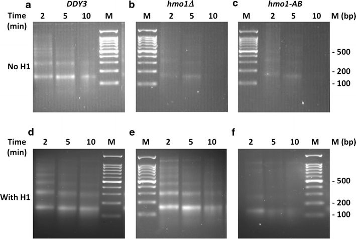
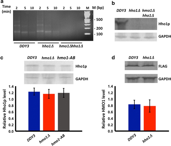
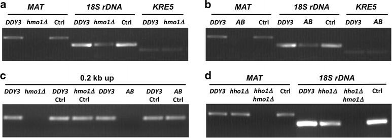
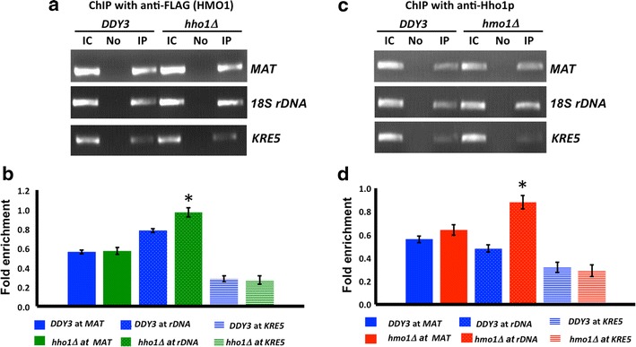
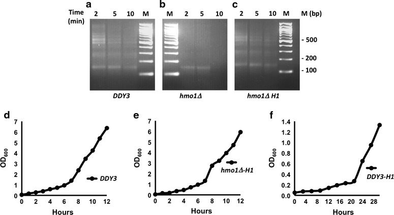
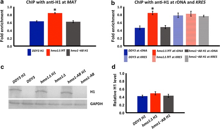
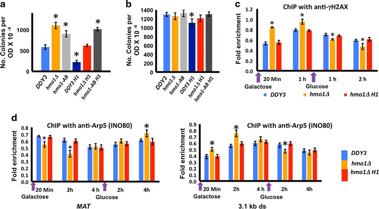
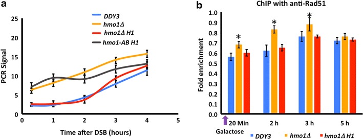
Results: We show here that HMO1 fulfills this function of a linker histone. For histone H1, chromatin compaction requires its basic C-terminal domain, and we find that the same pertains to HMO1, as deletion of its C-terminal lysine-rich extension renders chromatin nuclease sensitive. On rDNA, deletion of both HMO1 and Hho1p is required for significantly increased nuclease sensitivity. Expression of human histone H1 completely reverses the nuclease sensitivity characteristic of chromatin isolated from hmo1∆ cells. While chromatin remodeling events associated with repair of DNA double-strand breaks occur faster in the more dynamic chromatin environment created by the hmo1 deletion, expression of human histone H1 results in chromatin remodeling and double-strand break repair similar to that observed in wild-type cells.

Conclusion: Our data suggest that S. cerevisiae HMO1 protects linker DNA from nuclease digestion, a property also characteristic of mammalian linker histone H1. Notably, association with HMO1 creates a less dynamic chromatin environment that depends on its lysine-rich domain. That HMO1 has linker histone function has implications for investigations of chromatin structure and function as well as for evolution of proteins with roles in chromatin compaction.

Keywords: Chromatin; Chromatin immunoprecipitation; Double-strand break repair; Hho1p; High mobility group protein; Histone H1.


Figures

Fig. 1
Fig. 1
Resistance of chromatin to nuclease digestion requires linker histone H1 or HMO1 containing its lysine-rich extension. ac MNase digestion of chromatin isolated from wild-type cells (DDY3), hmo1∆, and hmo1-AB, respectively. df MNase digestion of chromatin isolated from wild-type, hmo1∆, and hmo1-AB cells expressing human linker histone H1.2 under control of a strong, constitutive promoter. Nuclei were digested with 0.25 U/µl MNase for the time indicated. Nucleosomal DNA was purified and resolved by agarose gel electrophoresis and stained with ethidium bromide
Fig. 2
Fig. 2
Effect of Hho1p on resistance of chromatin to nuclease digestion and on cellular content of HMO1. a MNase digestion of chromatin isolated from DDY3, hho1∆, and hmo1∆ hho1∆, respectively. Nuclei were digested with 0.25 U/µl MNase for the time indicated. Nucleosomal DNA was purified and resolved by agarose gel electrophoresis and stained with ethidium bromide. b Western blot of lysates from DDY3, hho1∆, and hmo1∆ hho1∆ using antibody to Hho1p or GAPDH. GAPDH migrates with a Mw ~36 kDa, while Hho1p migrates with a Mw ~28 kDa. c Western blot of lysates from DDY3, hmo1∆, and hmo1-ΑΒ using antibody to Hho1p or GAPDH. Densitometric analysis of three separate blots from three independent experiments shown below. Relative level = Hho1p/GAPDH. d Western blot of lysates from DDY3 and hho1∆ using antibody to FLAG-tagged HMO1 or GAPDH. HMO1-FLAG migrates with a Mw ~35 kDa. Densitometric analysis of three separate blots from three independent experiments shown below. Relative level = FLAG/GAPDH. Error bars represent standard deviation
Fig. 3
Fig. 3
Resistance of chromatin to MNase digestion monitored at specific loci. a, b Amplification of DNA representing MAT, 18S rDNA, and KRE5 after MNase digestion of chromatin isolated from wild-type cells (DDY3) and hmo1∆ (a) or hmo1-AB (b). c Amplification of DNA using primers annealing 0.2 kb upstream of the HO cleavage site within the MAT locus from DDY3, hmo1∆, and hmo1-AB. d Amplification of DNA representing MAT and 18S rDNA from chromatin isolated from DDY3, hho1∆, and hmo1∆hho1∆. In all panels, Ctrl denotes chromatin from the identified strain not incubated with MNase. Data are representative of three repeats
Fig. 4
Fig. 4
Effect of HMO1 or Hho1p on binding of the other protein. a Chromatin immunoprecipitation (ChIP) with DDY3 and hho1∆ using antibody to FLAG-tagged HMO1, monitoring binding at MAT, 18S rDNA, and KRE5. IC, input control; No, no antibody; IP, immunoprecipitation with anti-FLAG. b qRT-PCR analysis of ChIP data corresponding to (a). c ChIP with DDY3 and hmo1∆ using antibody to Hho1p, monitoring binding at MAT, 18S rDNA, and KRE5. d qRT-PCR analysis of ChIP data corresponding to (c). Data were normalized to corresponding input control at each time point. Fold enrichment = ChIP/Input DNA. Three independent experiments were performed. Error bars represent standard deviation. Asterisks represent statistical significance from DDY3 at the same locus based on Student’s t test (P < 0.05)
Fig. 5
Fig. 5
Equivalent binding of HMO1 to MAT in different growth phases. a, b Quantification by qRT-PCR of ChIP using antibody to FLAG-tagged HMO1 in DDY3, monitoring binding at the MAT locus. Data were normalized to corresponding input control. Error bars represent standard deviation of three experiments. a Cells recovering from quiescence (Rec) compared to stationary phase (Stat). b Cells in exponential phase (Exp) compared to stationary phase (Stat). Fold enrichment = ChIP/Input DNA. Three independent experiments were performed. Error bars represent standard deviation. c Growth of cells after inoculation of fresh media with stationary phase cells to OD600 ~0.05; cells were harvested for ChIP after 1 h (Rec) or 4 h (Exp)
Fig. 6
Fig. 6
Effect of linker histone H1 on MNase sensitivity of chromatin isolated from synchronized cells and on growth rate. ac MNase digestion of chromatin isolated from synchronized DDY3, hmo1∆, and hmo1∆ expressing human linker histone H1.2. Cells were synchronized in G1 phase for a total of 3 h by the addition of alpha factor. Nuclei were digested with 0.25 U/µl MNase for the time indicated. Nucleosomal DNA was purified and resolved by agarose gel electrophoresis and stained with ethidium bromide. df Growth curve for wild-type DDY3, hmo1∆ expressing H1, and DDY3 expressing H1. Cells were grown in synthetic-defined media, and cells were collected at regular intervals to measure OD at 600 nm
Fig. 7
Fig. 7
Both HMO1 and HMO1 deleted for its C-terminal tail compete with H1 for binding to chromatin. a Quantification by qRT-PCR of ChIP using antibody to H1 with DDY3, hmo1∆, and hmo1-AB strains, monitoring binding at the MAT locus. b qRT-PCR analysis of ChIP using antibody to H1, monitoring binding at 18S rDNA and at KRE5. Data were normalized to corresponding input control at each time point. Three independent experiments were performed. Error bars represent standard deviation. Asterisks represent statistical significance from DDY3 based on Student’s t test (P < 0.05). c Western blot using antibody to H1 showing equal protein level of histone H1 after transforming plasmid expressing human H1 under control of a strong, constitutive promoter in DDY3 (DDY3 H1), hmo1∆ (hmo1∆ H1), and hmo1-AB strain (hmo1-AB H1). Non-transformed cells DDY3, hmo1∆, and hmo1-AB were used as negative control. GAPDH expression levels were assessed in all samples as internal loading control, and the blots are representative of four independent experiments. GAPDH migrates with a Mw ~36 kDa, while H1 migrates with a Mw ~30 kDa (slower than its calculated Mw ~22 kDa). d Densitometric analysis of three separate blots from three independent experiments shown in (c). Relative H1 level = H1/GAPDH. Error bars represent standard deviation
Fig. 8
Fig. 8
Dynamic chromatin environment in hmo1∆ that leads to faster chromatin remodeling and DSB repair is restored to wild-type levels by expression of H1. a Survival of DDY3, hmo1∆, hmo1-AB and the corresponding strains expressing H1. After DSB induction, cells were plated and colonies counted. Three independent experiments were performed. Error bars represent standard deviation. b Cells not induced to express HO were plated as control. c qRT-PCR analysis of ChIP using antibody to phosphorylated H2A, monitoring presence of γ-H2AX at MAT during DNA damage (galactose) and repair (glucose). Data are normalized to corresponding input control at each time point. d qRT-PCR analysis of ChIP using antibody to Arp5, monitoring presence at MAT (left panel) and 3.1 kb downstream of DSB (right panel) during DNA damage (galactose) and repair (glucose). Data are normalized to corresponding input control at each time point. Three independent experiments were performed. Error bars represent standard deviation. In all panels, asterisks represent statistical significance from DDY3 at the respective time points based on Student’s t test (P < 0.05)
Fig. 9
Fig. 9
Chromatin state in hmo1∆ that leads to faster DNA end resection and faster Rad51 recruitment after DNA double-strand break is reversed on expression of H1. a Quantification of DNA resection by qRT-PCR using primers that anneal 1.6 kb upstream of the DSB. PCR products were amplified after exonuclease I treatment of genomic DNA isolated at the indicated times following DSB induction. All values were normalized to that for an independent locus (POL5). b qRT-PCR analysis of ChIP using antibody to Rad51, monitoring binding at MAT after DSB induction (galactose). Data are normalized to corresponding input control at each time point. Asterisks represent statistical significance from DDY3 at the respective time points based on Student’s t test (P < 0.05). Three independent experiments were performed

Similar articles

Cited by

References

    1. Hergeth SP, Schneider R. The H1 linker histones: multifunctional proteins beyond the nucleosomal core particle. EMBO Rep. 2015;16(11):1439–1453. doi: 10.15252/embr.201540749. - DOI - PMC - PubMed
    1. Harshman SW, Young NL, Parthun MR, Freitas MA. H1 histones: current perspectives and challenges. Nucleic Acids Res. 2013;41(21):9593–9609. doi: 10.1093/nar/gkt700. - DOI - PMC - PubMed
    1. Allan J, Mitchell T, Harborne N, Bohm L, Crane-Robinson C. Roles of H1 domains in determining higher order chromatin structure and H1 location. J Mol Biol. 1986;187(4):591–601. doi: 10.1016/0022-2836(86)90337-2. - DOI - PubMed
    1. Ellen TP, van Holde KE. Linker histone interaction shows divalent character with both supercoiled and linear DNA. Biochemistry. 2004;43(24):7867–7872. doi: 10.1021/bi0497704. - DOI - PubMed
    1. Lu X, Hamkalo B, Parseghian MH, Hansen JC. Chromatin condensing functions of the linker histone C-terminal domain are mediated by specific amino acid composition and intrinsic protein disorder. Biochemistry. 2009;48(1):164–172. doi: 10.1021/bi801636y. - DOI - PMC - PubMed

LinkOut - more resources