

Can Xanthophyll-Membrane Interactions Explain Their Selective Presence in the Retina and Brain?
- PMID: 27030822
- PMCID: PMC4809277
- DOI: 10.3390/foods5010007
Can Xanthophyll-Membrane Interactions Explain Their Selective Presence in the Retina and Brain?
Abstract
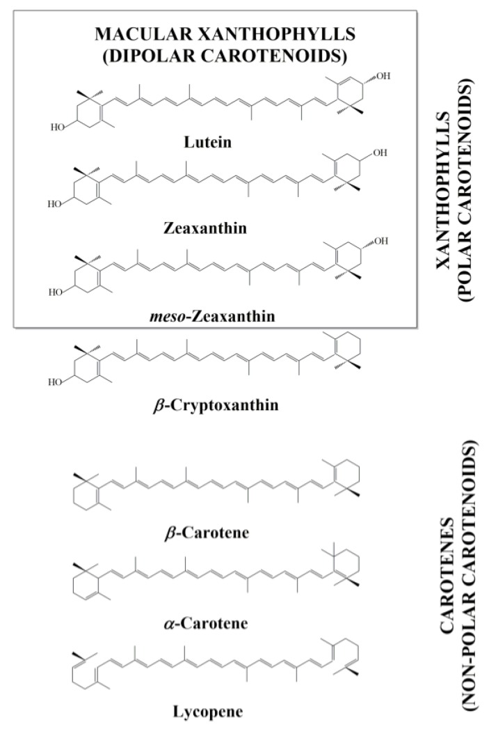
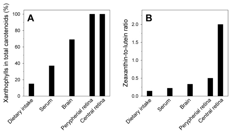
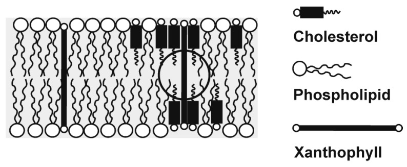
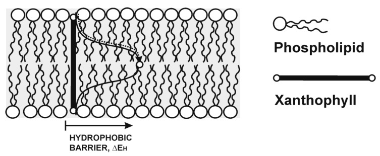
Epidemiological studies demonstrate that a high dietary intake of carotenoids may offer protection against age-related macular degeneration, cancer and cardiovascular and neurodegenerative diseases. Humans cannot synthesize carotenoids and depend on their dietary intake. Major carotenoids that have been found in human plasma can be divided into two groups, carotenes (nonpolar molecules, such as β-carotene, α-carotene or lycopene) and xanthophylls (polar carotenoids that include an oxygen atom in their structure, such as lutein, zeaxanthin and β-cryptoxanthin). Only two dietary carotenoids, namely lutein and zeaxanthin (macular xanthophylls), are selectively accumulated in the human retina. A third carotenoid, meso-zeaxanthin, is formed directly in the human retina from lutein. Additionally, xanthophylls account for about 70% of total carotenoids in all brain regions. Some specific properties of these polar carotenoids must explain why they, among other available carotenoids, were selected during evolution to protect the retina and brain. It is also likely that the selective uptake and deposition of macular xanthophylls in the retina and brain are enhanced by specific xanthophyll-binding proteins. We hypothesize that the high membrane solubility and preferential transmembrane orientation of macular xanthophylls distinguish them from other dietary carotenoids, enhance their chemical and physical stability in retina and brain membranes and maximize their protective action in these organs. Most importantly, xanthophylls are selectively concentrated in the most vulnerable regions of lipid bilayer membranes enriched in polyunsaturated lipids. This localization is ideal if macular xanthophylls are to act as lipid-soluble antioxidants, which is the most accepted mechanism through which lutein and zeaxanthin protect neural tissue against degenerative diseases.
Keywords: age-related macular degeneration (AMD); age-related neurodegenerative diseases; carotenoids; lipid antioxidants; lutein; macular xanthophylls; neural tissue; zeaxanthin.
Figures
Similar articles
-
Why has Nature Chosen Lutein and Zeaxanthin to Protect the Retina?J Clin Exp Ophthalmol. 2014 Feb 21;5(1):326. doi: 10.4172/2155-9570.1000326. J Clin Exp Ophthalmol. 2014. PMID: 24883226 Free PMC article.
-
Location of macular xanthophylls in the most vulnerable regions of photoreceptor outer-segment membranes.Arch Biochem Biophys. 2010 Dec 1;504(1):61-6. doi: 10.1016/j.abb.2010.05.015. Epub 2010 May 28. Arch Biochem Biophys. 2010. PMID: 20494651 Free PMC article. Review.
-
Why is Zeaxanthin the Most Concentrated Xanthophyll in the Central Fovea?Nutrients. 2020 May 7;12(5):1333. doi: 10.3390/nu12051333. Nutrients. 2020. PMID: 32392888 Free PMC article. Review.
-
Factors Differentiating the Antioxidant Activity of Macular Xanthophylls in the Human Eye Retina.Antioxidants (Basel). 2021 Apr 14;10(4):601. doi: 10.3390/antiox10040601. Antioxidants (Basel). 2021. PMID: 33919673 Free PMC article. Review.
-
Lutein, zeaxanthin, and the macular pigment.Arch Biochem Biophys. 2001 Jan 1;385(1):28-40. doi: 10.1006/abbi.2000.2171. Arch Biochem Biophys. 2001. PMID: 11361022 Review.
Cited by
-
Relation Between Dietary Carotenoid Intake, Serum Concentration, and Mortality Risk of CKD Patients Among US Adults: National Health and Nutrition Examination Survey 2001-2014.Front Med (Lausanne). 2022 Jul 8;9:871767. doi: 10.3389/fmed.2022.871767. eCollection 2022. Front Med (Lausanne). 2022. PMID: 35872751 Free PMC article.
-
Localization and Orientation of Xanthophylls in a Lipid Bilayer.Sci Rep. 2017 Aug 29;7(1):9619. doi: 10.1038/s41598-017-10183-7. Sci Rep. 2017. PMID: 28852075 Free PMC article.
-
Different Doses of β-Cryptoxanthin May Secure the Retina from Photooxidative Injury Resulted from Common LED Sources.Oxid Med Cell Longev. 2021 Feb 10;2021:6672525. doi: 10.1155/2021/6672525. eCollection 2021. Oxid Med Cell Longev. 2021. PMID: 33628377 Free PMC article.
-
Advancing nutrition science to meet evolving global health needs.Eur J Nutr. 2023 Dec;62(Suppl 1):1-16. doi: 10.1007/s00394-023-03276-9. Epub 2023 Nov 28. Eur J Nutr. 2023. PMID: 38015211 Free PMC article.
-
Xanthophyll pigments dietary supplements administration and retinal health in the context of increasing life expectancy trend.Front Nutr. 2023 Aug 10;10:1226686. doi: 10.3389/fnut.2023.1226686. eCollection 2023. Front Nutr. 2023. PMID: 37637949 Free PMC article.
References
-
- Johnson E.J., Vishwanathan R., Johnson M.A., Hausman D.B., Davey A., Scott T.M., Green R.C., Miller L.S., Gearing M., Woodard J., et al. Relationship between Serum and Brain Carotenoids, alpha-Tocopherol, and Retinol Concentrations and Cognitive Performance in the Oldest Old from the Georgia Centenarian Study. J. Aging Res. 2013;2013:951786. doi: 10.1155/2013/951786. - DOI - PMC - PubMed
-
- Craft N.E., Haitema T.B., Garnett K.M., Fitch K.A., Dorey C.K. Carotenoid, tocopherol, and retinol concentrations in elderly human brain. J. Nutr. Health Aging. 2004;8:156–162. - PubMed
-
- Bone R.A., Landrum J.T., Fernandez L., Tarsis S.L. Analysis of the macular pigment by HPLC: Retinal distribution and age study. Investig. Ophthalmol. Vis. Sci. 1988;29:843–849. - PubMed
-
- Snodderly D.M., Auran J.D., Delori F.C. The macular pigment. II. Spatial distribution in primate retinas. Investig. Ophthalmol. Vis. Sci. 1984;25:674–685. - PubMed
Grants and funding
LinkOut - more resources
Full Text Sources
Other Literature Sources
