

Lethal neonatal meningoencephalitis caused by multi-drug resistant, highly virulent Escherichia coli
- PMID: 27030919
- PMCID: PMC4818964
- DOI: 10.3109/23744235.2016.1144142
Lethal neonatal meningoencephalitis caused by multi-drug resistant, highly virulent Escherichia coli
Abstract
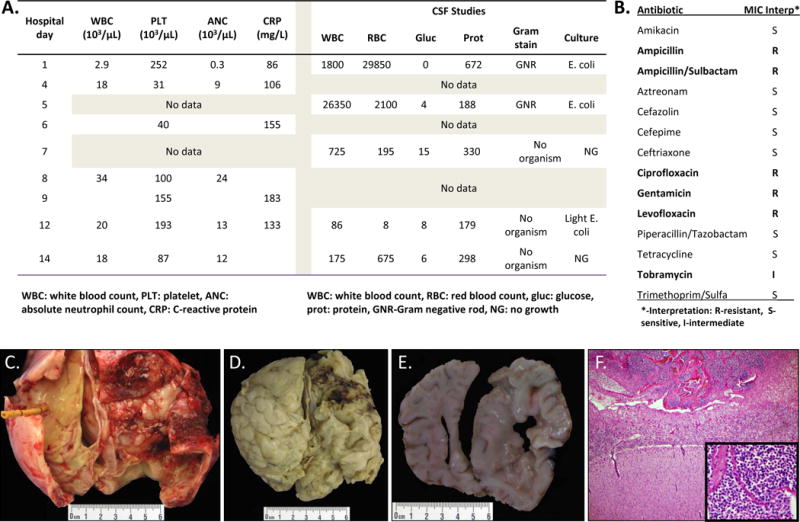
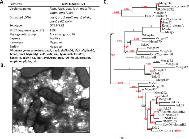
Neonatal meningitis is a rare but devastating condition. Multi-drug resistant (MDR) bacteria represent a substantial global health risk. This study reports on an aggressive case of lethal neonatal meningitis due to a MDR Escherichia coli (serotype O75:H5:K1). Serotyping, MDR pattern and phylogenetic typing revealed that this strain is an emergent and highly virulent neonatal meningitis E. coli isolate. The isolate was resistant to both ampicillin and gentamicin; antibiotics currently used for empiric neonatal sepsis treatment. The strain was also positive for multiple virulence genes including K1 capsule, fimbrial adhesion fimH, siderophore receptors iroN, fyuA and iutA, secreted autotransporter toxin sat, membrane associated proteases ompA and ompT, type II polysaccharide synthesis genes (kpsMTII) and pathogenicity-associated island (PAI)-associated malX gene. The presence of highly-virulent MDR organisms isolated in neonates underscores the need to implement rapid drug resistance diagnostic methods and should prompt consideration of alternate empiric therapy in neonates with Gram negative meningitis.
Keywords: Escherichia coli; hypervirulence; meningitis; meningoencephalitis; multi-drug resistance; neonate.
Conflict of interest statement
This work was funded in part by the Department of Pediatrics, Vanderbilt University School of Medicine to O.G.G-D; NIH K08GM106143,
Figures
Similar articles
-
Case report: A rare multidrug-resistant Escherichia coli causes fatal neonatal meningoencephalitis.Front Public Health. 2023 Jul 28;11:1174536. doi: 10.3389/fpubh.2023.1174536. eCollection 2023. Front Public Health. 2023. PMID: 37575122 Free PMC article.
-
Bacterial characteristics of importance for recurrent urinary tract infections caused by Escherichia coli.Dan Med Bull. 2011 Apr;58(4):B4187. Dan Med Bull. 2011. PMID: 21466767 Review.
-
Outbreak Caused by Escherichia coli O18: K1: H7 Sequence Type 95 in a Neonatal Intensive Care Unit in Barcelona, Spain.Pediatr Infect Dis J. 2017 Nov;36(11):1079-1086. doi: 10.1097/INF.0000000000001652. Pediatr Infect Dis J. 2017. PMID: 28650938
-
Antimicrobial susceptibility of 136 Escherichia coli isolates from cases of neonatal meningitis and relationship with virulence.Clin Microbiol Infect. 2007 Dec;13(12):1207-10. doi: 10.1111/j.1469-0691.2007.01838.x. Epub 2007 Oct 19. Clin Microbiol Infect. 2007. PMID: 17949439
-
E. coli meningitis: K1 antigen and virulence.Annu Rev Med. 1978;29:129-36. doi: 10.1146/annurev.me.29.020178.001021. Annu Rev Med. 1978. PMID: 348030 Review.
Cited by
-
Case Report: Fatal Outcome for a Preterm Newborn With Meningitis Caused by Extended-Spectrum β-Lactamase-Producing Escherichia coli Sequence Type 1193.Front Pediatr. 2022 Apr 6;10:866762. doi: 10.3389/fped.2022.866762. eCollection 2022. Front Pediatr. 2022. PMID: 35463903 Free PMC article.
-
The complete genome sequence of five pre-2013 Escherichia coli sequence type (ST)1193 strains reveals insights into an emerging pathogen.Access Microbiol. 2024 Oct 18;6(10):000894.v3. doi: 10.1099/acmi.0.000894.v3. eCollection 2024. Access Microbiol. 2024. PMID: 39430659 Free PMC article.
-
Escherichia coli K1 Modulates Peroxisome Proliferator-Activated Receptor γ and Glucose Transporter 1 at the Blood-Brain Barrier in Neonatal Meningitis.J Infect Dis. 2016 Oct 1;214(7):1092-104. doi: 10.1093/infdis/jiw306. Epub 2016 Jul 24. J Infect Dis. 2016. PMID: 27456707 Free PMC article.
-
Septic shock due to Escherichia coli meningoencephalitis treated with immunoglobulin-M-enriched immunoglobulin preparation as adjuvant therapy: a case report.J Med Case Rep. 2021 Mar 29;15(1):138. doi: 10.1186/s13256-021-02731-7. J Med Case Rep. 2021. PMID: 33775244 Free PMC article.
-
Prevalence of Escherichia coli ST1193 Causing Intracranial Infection in Changsha, China.Trop Med Infect Dis. 2022 Aug 31;7(9):217. doi: 10.3390/tropicalmed7090217. Trop Med Infect Dis. 2022. PMID: 36136628 Free PMC article.
References
-
- Barichello T, Dagostim VS, Generoso JS, Simoes LR, Dominguini D, Silvestre C, et al. Neonatal Escherichia coli K1 meningitis causes learning and memory impairments in adulthood. J Neuroimmunol. 2014;272:35–41. - PubMed
Publication types
MeSH terms
Substances
Grants and funding
LinkOut - more resources
Full Text Sources
Other Literature Sources
Medical
Molecular Biology Databases
Research Materials