. 2016 Mar 31;12(3):e1005508.
doi: 10.1371/journal.ppat.1005508. eCollection 2016 Mar.

PI3Kγ Is Critical for Dendritic Cell-Mediated CD8+ T Cell Priming and Viral Clearance during Influenza Virus Infection

Affiliations

PI3Kγ Is Critical for Dendritic Cell-Mediated CD8+ T Cell Priming and Viral Clearance during Influenza Virus Infection

Samuel Philip Nobs et al. PLoS Pathog. .

Abstract

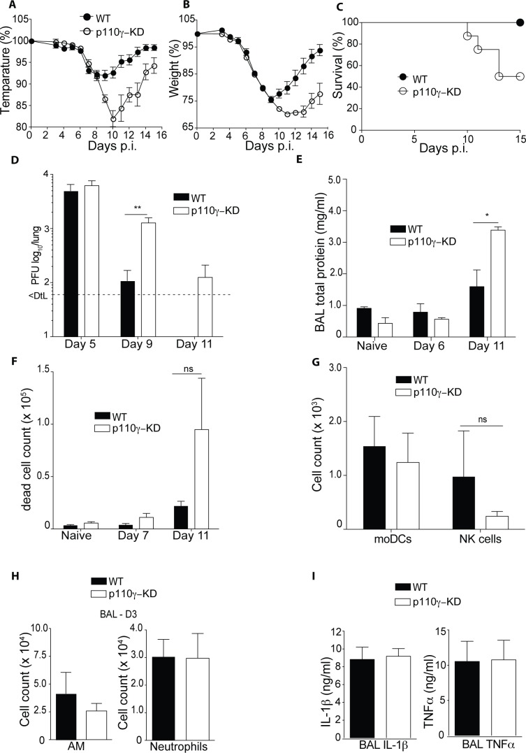
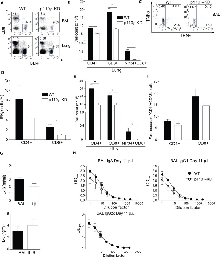
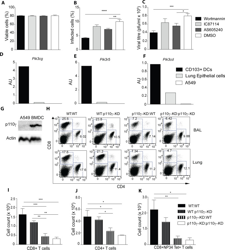
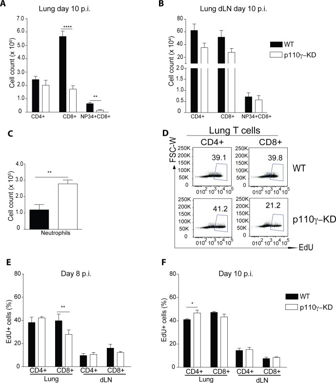
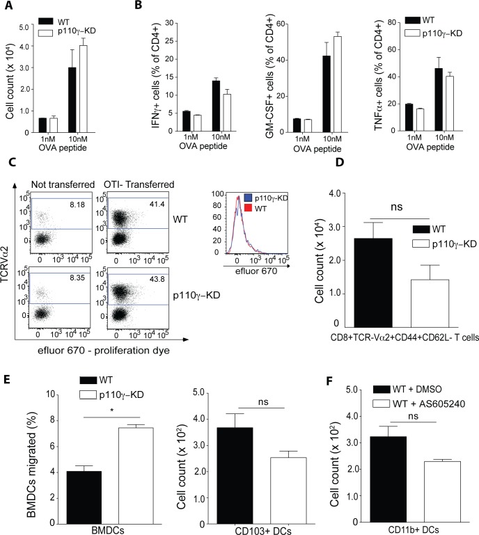
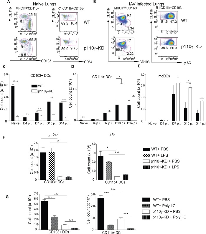
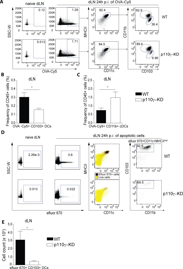
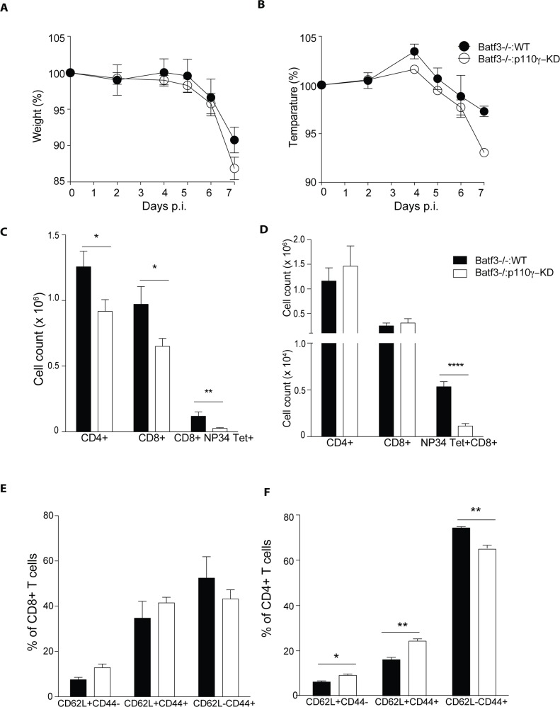
Phosphoinositide-3-kinases have been shown to be involved in influenza virus pathogenesis. They are targeted directly by virus proteins and are essential for efficient viral replication in infected lung epithelial cells. However, to date the role of PI3K signaling in influenza infection in vivo has not been thoroughly addressed. Here we show that one of the PI3K subunits, p110γ, is in fact critically required for mediating the host's antiviral response. PI3Kγ deficient animals exhibit a delayed viral clearance and increased morbidity during respiratory infection with influenza virus. We demonstrate that p110γ is required for the generation and maintenance of potent antiviral CD8+ T cell responses through the developmental regulation of pulmonary cross-presenting CD103+ dendritic cells under homeostatic and inflammatory conditions. The defect in lung dendritic cells leads to deficient CD8+ T cell priming, which is associated with higher viral titers and more severe disease course during the infection. We thus identify PI3Kγ as a novel key host protective factor in influenza virus infection and shed light on an unappreciated layer of complexity concerning the role of PI3K signaling in this context.


Conflict of interest statement

The authors have declared that no competing interests exist.

Figures

Fig 1
Fig 1. p110γ-deficiency is associated with severe disease and delayed viral clearance during influenza virus infection.
WT and p110γ-KD mice were infected with 50 pfu PR8 IAV, shown are (A) temperature and (B) weight, relative to the day of infection (n = 8). WT and p110γ-KD mice were infected with 200 pfu, shown is(C) a survival curve showing the fraction of surviving animals for each day p.i (n = 10). WT and p110γ-KD mice were infected with 50 pfu PR8 IAV, shown are (D) lung viral titers (n = 5), (E) BAL total protein content (n = 5) and (F) number of eFluor780+ dead cells in the BAL of infected animals at different time-points (n = 5). At day 3 p.i. the immune cell infilitrate in the BAL was quantified, all cells were pregated on CD45+ viable cells, shown are (G) moDCs identified as CD11c+MHCII+CD11b+Ly-6C+ cells, NK cells identified as CD49b+CD3-NK1.1+ cells, (H) AM, identified as Siglec-F+ CD11c+ cells and neutrophils as CD11b+Ly-6G+ cells (n = 4) (I) At day 3 p.i. the cytokine levels of TNFα and IL-1β in the BAL was quantified using ELISA (n = 4). For all bar graphs mean ± SEM is shown and results are representative of at least 2 experiments. The Student’s t test (unpaired) was used: p < 0.05 (*), p < 0.01 (**), p < 0.001 (***), p < 0.0001 (****).
Fig 2
Fig 2. p110γ is required for the antiviral CD8+ T cell response.
WT and p110γ-KD mice were infected with 50 pfu PR8 IAV and the T cell response was analysed at day 7 p.i. Cells were pregated on CD45+ viable cells. Shown are (A-B) representative dot plots of CD4+ and CD8+ T cells in the BAL and lungs of infected animals as well as (B) a summary of the lungs of all mice including NP34 Tet+ virus-specific CD8+ T cells (mean ± SEM) (n = 5) (C-D) BAL cells from infected animals were restimulated with NP34 peptide and inactivated PR8 IAV, shown are representative dot plots as a well as a summary of all animals (n = 5) (mean ± SEM). (E) Shown is a quantification of T cells of the lung dLN of all analysed mice (mean ± SEM)(F) Activated T cells were identified as CD44+CD62L-. Shown are the fold increase in total numbers compared to naive lungs (G) Shown is a summary of the cytokine levels in the BAL of infected animals (mean ± SEM) (n = 5). (H) Antibody titres of virus-specific antibodies in the BAL were analysed at day 11 p.i., shown is a titration curve of IgA, IgG1 and IgG2c isotypes (mean ± SEM) (n = 5). Results are representative of at least 2 experiments. The Student’s t test (unpaired) was used: p < 0.05 (*), p < 0.01 (**), p < 0.001 (***), p < 0.0001 (****).
Fig 3
Fig 3. The antiviral T cell response is dependent on p110γ in the hematopoietic compartment.
(A-C) A549 cells were treated with inhibitors against p110γ (AS650240), p110d (IC87114), all PI3K subunits (Wortmannin) or DMSO as a control for 30 mins before infecting with PR8 IAV MOI 0.5. Shown is the frequency of viable cells (A) infected cells (B) and the viral titre (C) after 10h of infection (n = 6) (D-F) CD103+ DCs and lung epithelial cells were sorted from WT animals and expression of different PI3K subunits was measured by qPCR, A549 cells were also included. Shown is the expression of Pik3cg, Pik3r5 and Pik3cd normalized to Tbp. Lung CD103+ DCs were identified as CD45+SiglecF-CD11c+MHCII+CD103+CD11b- and lung epithelial cells as CD45-podoplanin+ cells respectively (G) Shown is a western blot of A549 and bone-marrow derived dendritic cells (BMDCs) of p110γ (H-K) WT→WT, WT→ p110γ-KD, p110γ-KD→WT and p110γ-KD→p110γ-KD BM chimeras were generated and subsequently infected with 50 pfu PR8 IAV. The T cell response was analyzed at day 7 p.i. Shown are representative dot plots of CD4+ and CD8+ T cells as well as a summary of all infected animals including NP34 Tet+ CD8+ T cells (mean ± SEM) (n = 5). The results are representative of 2 experiments. One way ANOVA was used with CI 95%: p < 0.05 (*), p < 0.01 (**), p < 0.001 (***), p < 0.0001 (****).
Fig 4
Fig 4. p110γ is largely dispensable for T cell proliferation during IAV infection.
WT and p110γ-KD mice were infected with 50 pfu PR8 IAV and the immune cell infiltrate was analysed at day 10 p.i. Cells were pregated on CD45+ viable cells. Shown are (A-B) a summary of T cells in lungs and lung dLN of all mice including NP34 Tet+ CD8+ T cells (mean ± SEM) as well as (C) Neutrophils in the lungs of infected animals (mean ± SEM) (n = 6). At day 6 and day 7 p.i. mice were injected i.p. with EdU and lungs and dLN of infected animals were analyzed on day 8 p.i. to evaluate frequencies of EdU+ cells. Shown are (D) representative dot plots of lung T cells (E) as well as a summary of all infected animals (mean ± SEM) (n = 6). (F) At day 8 and day 9 p.i. mice were injected i.p. with EdU and lungs and dLN of infected animals were analyzed on day 10 p.i. to evaluate frequencies of EdU+ cells. Shown is summary of all infected animals in the lungs and lung dLN (mean ± SEM) (n = 6). The Student’s t test (unpaired) was used: p < 0.05 (*), p < 0.01 (**), p < 0.001 (***), p < 0.0001 (****).
Fig 5
Fig 5. p110γ is dispensable for DC-induced T cell polarization and migration in vitro.
WT and p110γ-KD BMDCs were cocultured with OTII CD4+ T cells sorted from the spleen and different concentrations of OVA323-339 peptide for 4 days. Shown are (A) total cell counts of CD4+ T cells (B) as well as production of IFNγ, GM-CSF and TNFα after 4h PMA/ionomcyin restimulation (n = 3). (C-D) 5x10⁶ OT-I cells labeled with efluor 670 were transferred into WT and p110g-KD recipients at day 0. On day 1 after transfer mice received 4mg OVA/Alum into the flank. At day 7p.i. the inguinal lymph nodes were analysed. (C) Shown are representative dot plots of recipients and non-transferred controls as well as a representative histogram overlay of transferred WT and A 6h transwell assay with 500ng/ml CCL21 was conducted with either (C) BMDCs of WT and p110g-KD mice. (D) Shown is the total number of CD8+TCRVa2+CD44+CD62L cells in the inguinal lymph nodes of all analysed animals (mean ± SEM) (n = 5). (E-F) A 6h transwell assay with 500ng/ml CCL21 was conducted with either (E) BMDCs of WT or p110γ-KD origin or (n = 2) (F) dendritic cell subsets sorted from naïve lungs of WT mice (n = 3). (C) Shown is the frequency of migrated cells as summary of all wells (mean ± SEM) (C) or the total number of cells which migrated respectively (D). Lung dendritic cells were sorted as either CD45+Siglec-F-CD11c+MHCII+CD103+CD11b- or CD45+Siglec-F-CD11c+MHCII+CD103-CD11b+CD64- cells representing CD103+ and CD11b+ conventional DCs respectively. Lung DCs were incubated with either 15 μM AS605240 or DMSO as a control. For A-B Ooe way ANOVA was used with CI 95%h. For C-F the student’s t test (unpaired) was used: p < 0.05 (*), p < 0.01 (**), p < 0.001 (***), p < 0.0001 (****).
Fig 6
Fig 6. p110γ is essential for lung DC development in homeostasis and respiratory viral infection.
WT and p110γ-KD mice were infected with 50 pfu PR8 IAV and the dendritic cell compartment in the lung and BAL was analysed at day 7 and day 10 p.i. Naïve animals are included as a comparison. Shown are (A-B) representative dot plots of naïve and infected lungs as well (C-E) as a summary of infected animals for CD103+ DCs, CD11b+ DCs and moDCs in the lung (n = 4–6) (mean ± SEM). Cells were pregated as CD45+SiglecF-CD11c+MHCII+ viable cells. CD103+ DCs were identified in naïve and infected lungs as CD103+CD11b- cells. CD11b+ DCs were identified as CD11b+CD64- in naïve and CD11b+Ly-6C- in infected lungs and moDCs were identified as CD11b+Ly-6C+ cells in infected lungs and BAL. (F) WT and p110γ-KD mice were inoculated i.t. with 100ng LPS or PBS as a control. Lungs were harvested at 24h or 48h post injection and cellular composition was analysed using flow cytometry. Shown is a summary of FACS dot plots showing the total number of CD103+ dendritic cells (mean ± SEM) (n = 3). (G) WT and p110γ-KD mice were inoculated i.t. with 50ug Poly I:C or PBS as a control. Lungs were harvested at 24h post injection and cellular composition was analysed using flow cytometry. Shown are a summary of FACS dot plots showing the total number of lung CD103+ and CD11b+ dendritic cell subsets (mean ± SEM) (n = 4). The data is representative of at least 2 experiments. The Student’s t test (unpaired) was used: p < 0.05 (*), p < 0.01 (**), p < 0.001 (***), p < 0.0001 (****).
Fig 7
Fig 7. p110γ is required for lung CD103+ DC-mediated transport of antigen to the dLN.
(A-C) WT and p110γ-KD mice were injected intratracheally with 40μg OVA-Cy5 and 100ng LPS and sacrificed 24h later for analysis of the lung and the dLN by flow cytometry. DC subsets were identified as CD45+CD11c+Siglec F-MHCIIhigh cells, shown are (A) representative FACS dot plots, as well as a summary of OVA-Cy5+ CD103+ DCs (B) and CD11b+ DCs (C) in the dLN (mean ± SEM) (n = 4), (D-E) p110γ-KD mice were inoculated i.t. with efluor 670 -labeled apoptotic thymocytes or no cells as a control. Lung dLN cells were isolated 24 h later and analyzed by flow cytometry. Efferocytic cells were identified as efluor 670+, and migratory DCs were identified as CD11c+MHCIIhigh. (D) Overlays show efluor 670+ cells (black) and total live cells (yellow) as indicated. (E) Total numbers of efluor 670+ DCs that express CD103 are shown (mean ± SEM) (n = 4). The data is representative of at least 2 experiments. The Student’s t test (unpaired) was used: p < 0.05 (*), p < 0.01 (**), p < 0.001 (***), p < 0.0001 (****).
Fig 8
Fig 8. p110γ in lung CD103+ DCs is required for the antiviral CD8+ T cell response.
WT mice were irradiated and reconstituted with either Batf3-/-:WT or Batf3-/-:p110γ-KD BM at a ratio of 4:1. 10 weeks after reconstitution mice were infected with 50 pfu PR8 IAV. Shown are (A) the weight and (B) temperature loss during the course of infection. The antiviral T cell response was analysed at day 7 p.i. Shown are a summary FACS dot plots of analysed organs, including the lungs (C) and the lung dLN (D) (mean ± SEM) (n = 5) (E-F) The frequency of CD62L and CD44 expressing T cells in the lungs of infected animals. (mean ± SEM) (n = 5) for all graphs. The Student’s t test (unpaired) was used: p < 0.05 (*), p < 0.01 (**), p < 0.001 (***), p < 0.0001 (****).

Similar articles

Cited by

References

    1. Zvelebil MJ, MacDougall L, Leevers S, Volinia S, Vanhaesebroeck B, Gout I, et al. Structural and functional diversity of phosphoinositide 3-kinases. Philosophical transactions of the Royal Society of London Series B, Biological sciences. 1996;351(1336):217–23. 10.1098/rstb.1996.0019 . - DOI - PubMed
    1. Hiles ID, Otsu M, Volinia S, Fry MJ, Gout I, Dhand R, et al. Phosphatidylinositol 3-kinase: structure and expression of the 110 kd catalytic subunit. Cell. 1992;70(3):419–29. . - PubMed
    1. Hu P, Mondino A, Skolnik EY, Schlessinger J. Cloning of a novel, ubiquitously expressed human phosphatidylinositol 3-kinase and identification of its binding site on p85. Mol Cell Biol. 1993;13(12):7677–88. . - PMC - PubMed
    1. Vanhaesebroeck B, Welham MJ, Kotani K, Stein R, Warne PH, Zvelebil MJ, et al. P110delta, a novel phosphoinositide 3-kinase in leukocytes. Proc Natl Acad Sci U S A. 1997;94(9):4330–5. . - PMC - PubMed
    1. Otsu M, Hiles I, Gout I, Fry MJ, Ruiz-Larrea F, Panayotou G, et al. Characterization of two 85 kd proteins that associate with receptor tyrosine kinases, middle-T/pp60c-src complexes, and PI3-kinase. Cell. 1991;65(1):91–104. . - PubMed

Publication types

MeSH terms

Substances

LinkOut - more resources