

Quantification of Ellipsoid Zone Changes in Retinitis Pigmentosa Using en Face Spectral Domain-Optical Coherence Tomography
- PMID: 27031504
- PMCID: PMC5317200
- DOI: 10.1001/jamaophthalmol.2016.0502
Quantification of Ellipsoid Zone Changes in Retinitis Pigmentosa Using en Face Spectral Domain-Optical Coherence Tomography
Erratum in
-
Incorrect Term in the Methods and Discussion.JAMA Ophthalmol. 2016 Jul 1;134(7):849. doi: 10.1001/jamaophthalmol.2016.2062. JAMA Ophthalmol. 2016. PMID: 27416273 No abstract available.
Abstract
Importance: New methods are needed to quantify the change in the outer retinal structures in retinitis pigmentosa (RP).
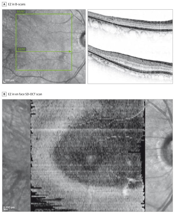
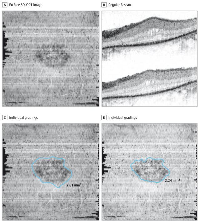
Objective: To implement an alternate method for tracking ellipsoid zone (EZ) changes in RP by quantifying the EZ area on en face spectral domain-optical coherence tomographic (SD-OCT) images.
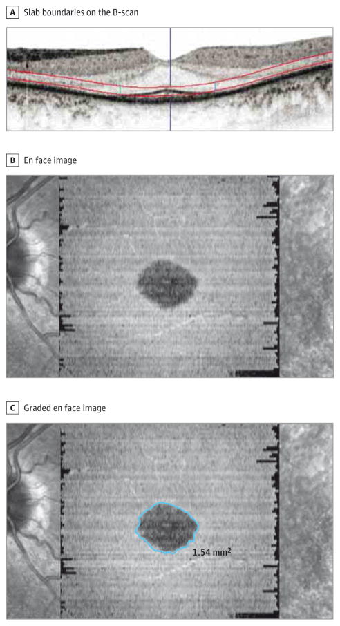
Design, setting, and participants: Data for this observational case study were collected at the Department of Ophthalmology, University of California, Los Angeles, from May 1 to July 30, 2015, and included SD-OCT images of a subset of patients from the Trial of Oral Valproic Acid for Retinitis Pigmentosa. To be eligible for the en face OCT subanalysis, the preserved EZ area was required to be limited to the SD-OCT scanning field. Cases in which the EZ band extended to the margins of any B-scan or the most superior or inferior B-scan were excluded. The SD-OCT images of all included cases were imported into the manufacturer's software to generate en face images at the level of the EZ. Two certified SD-OCT graders independently delineated the boundaries of the preserved EZ on the en face images.
Main outcomes and measures: Comparison of the 2 masked gradings of the generated en face images of patients with RP for agreement between the graders and the validity of the method.
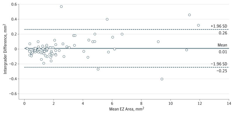
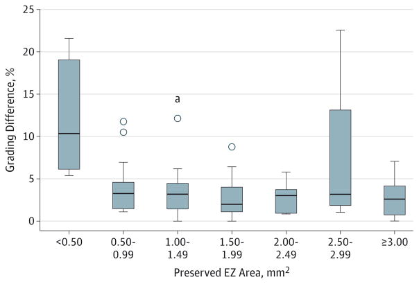
Results: Of the 43 available patients with volume SD-OCT data, 45 eyes of 24 patients met the eligibility criteria and were included in this subanalysis. Every patient had 2 visits that were 1 year apart, which included a total of 90 en face OCT images that were graded. The mean (SD) absolute difference and percentage difference between the 2 independent graders for each visit were 0.08 (0.10) mm2 and 4.5% (5.9%), respectively. The EZ area determined by the 2 graders showed excellent agreement with an intraclass correlation coefficient of 0.996 (95% CI, 0.995-0.997; P < .001).
Conclusions and relevance: Quantification of the preserved EZ area on en face SD-OCT images of patients with RP is a valid and reproducible method. En face SD-OCT quantification may be a useful tool for monitoring the EZ changes of patients with advanced RP and a useful outcome measurement variable in therapeutic trials.
Conflict of interest statement
Figures
Similar articles
-
Correlation between B-scan optical coherence tomography, en face thickness map ring and hyperautofluorescent ring in retinitis pigmentosa patients.Graefes Arch Clin Exp Ophthalmol. 2019 Aug;257(8):1601-1609. doi: 10.1007/s00417-019-04265-7. Epub 2019 May 2. Graefes Arch Clin Exp Ophthalmol. 2019. PMID: 31049658 Free PMC article.
-
Spectral-domain optical coherence tomography measures of outer segment layer progression in patients with X-linked retinitis pigmentosa.JAMA Ophthalmol. 2013 Sep;131(9):1143-50. doi: 10.1001/jamaophthalmol.2013.4160. JAMA Ophthalmol. 2013. PMID: 23828615 Free PMC article. Clinical Trial.
-
Correlating optical coherence tomography biomarkers with visual acuity in nigerian retinitis pigmentosa patients.Niger J Clin Pract. 2022 Mar;25(3):267-272. doi: 10.4103/njcp.njcp_1312_21. Niger J Clin Pract. 2022. PMID: 35295047
-
Optical Coherence Tomographic Analysis of Retina in Retinitis Pigmentosa Patients.Ophthalmic Res. 2016;56(3):111-22. doi: 10.1159/000445063. Epub 2016 Jun 29. Ophthalmic Res. 2016. PMID: 27352292 Review.
-
Natural history and clinical biomarkers of progression in X-linked retinitis pigmentosa: a systematic review.Acta Ophthalmol. 2021 Aug;99(5):499-510. doi: 10.1111/aos.14662. Epub 2020 Nov 30. Acta Ophthalmol. 2021. PMID: 33258268
Cited by
-
Deep Learning-Facilitated Study of the Rate of Change in Photoreceptor Outer Segment Metrics in RPGR-Related X-Linked Retinitis Pigmentosa.Invest Ophthalmol Vis Sci. 2023 Nov 1;64(14):31. doi: 10.1167/iovs.64.14.31. Invest Ophthalmol Vis Sci. 2023. PMID: 37988107 Free PMC article.
-
Widefield OCT angiography.Prog Retin Eye Res. 2025 Jul;107:101378. doi: 10.1016/j.preteyeres.2025.101378. Epub 2025 Jun 13. Prog Retin Eye Res. 2025. PMID: 40517946 Review.
-
Determination of Length of Interdigitation Zone by Optical Coherence Tomography and Retinal Sensitivity by Microperimetry and Their Relationship to Progression of Retinitis Pigmentosa.Biomed Res Int. 2019 Jun 20;2019:1217270. doi: 10.1155/2019/1217270. eCollection 2019. Biomed Res Int. 2019. PMID: 31321226 Free PMC article.
-
Retinal Imaging Findings in Inherited Retinal Diseases.J Clin Med. 2024 Apr 3;13(7):2079. doi: 10.3390/jcm13072079. J Clin Med. 2024. PMID: 38610844 Free PMC article. Review.
-
Structural evaluation in inherited retinal diseases.Br J Ophthalmol. 2021 Dec;105(12):1623-1631. doi: 10.1136/bjophthalmol-2021-319228. Epub 2021 May 12. Br J Ophthalmol. 2021. PMID: 33980508 Free PMC article. Review.
References
Publication types
MeSH terms
Substances
Grants and funding
LinkOut - more resources
Full Text Sources
Other Literature Sources
Medical
Research Materials
Miscellaneous
