. 2016 Mar 31;7(3):e2163.
doi: 10.1038/cddis.2016.14.

Renal cell carcinoma escapes death by p53 depletion through transglutaminase 2-chaperoned autophagy

Affiliations

Renal cell carcinoma escapes death by p53 depletion through transglutaminase 2-chaperoned autophagy

J H Kang et al. Cell Death Dis. .

Abstract

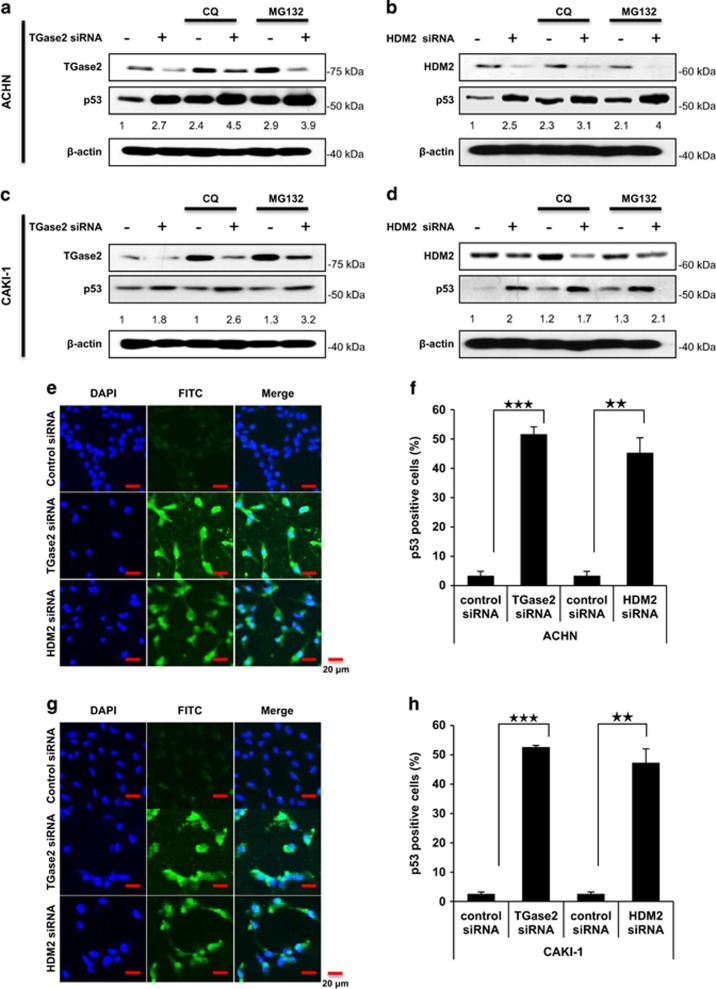
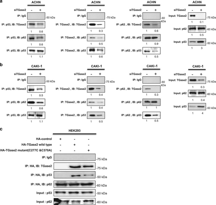
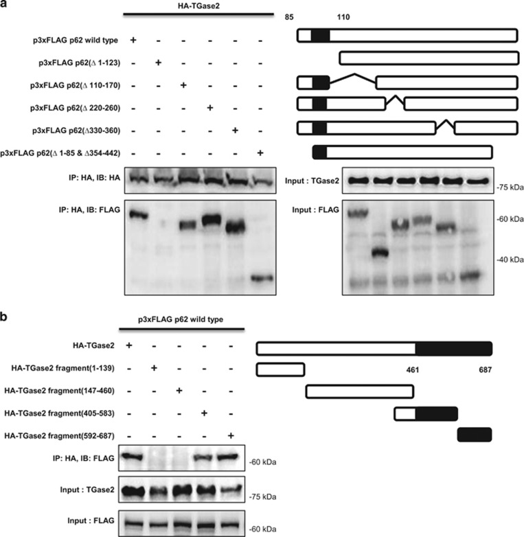
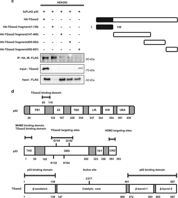
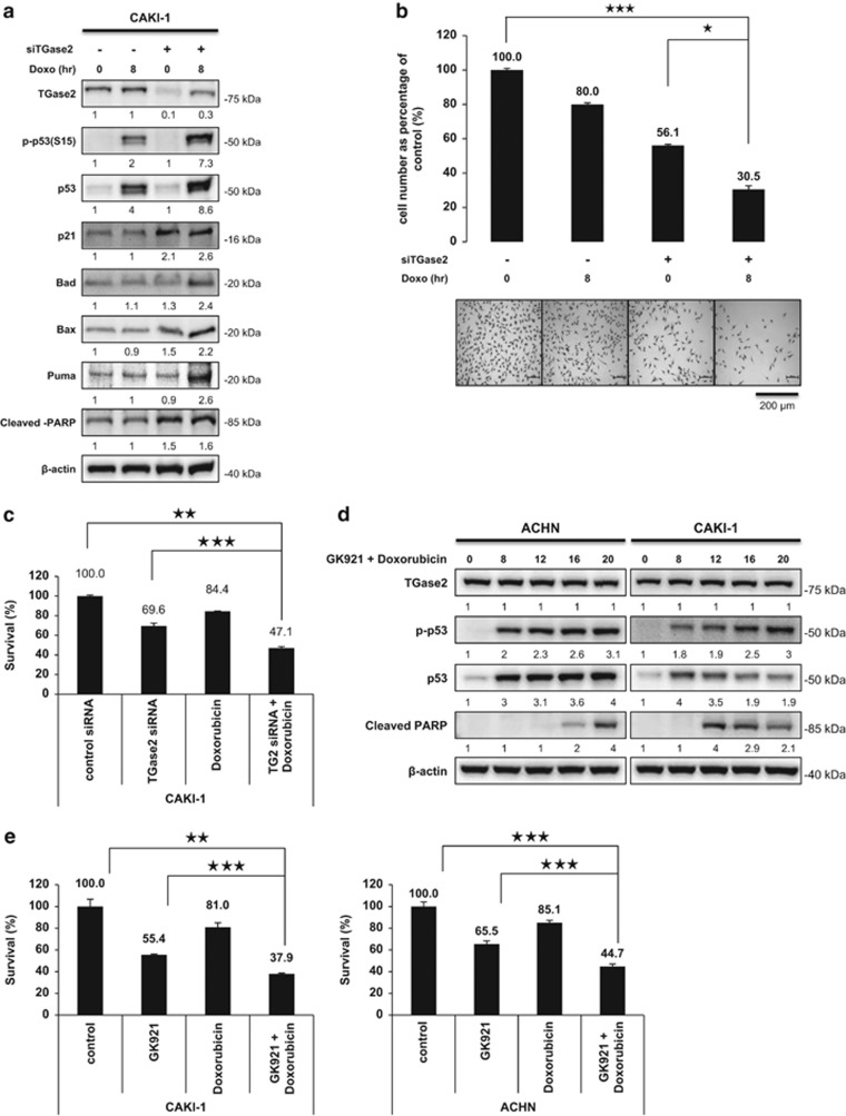
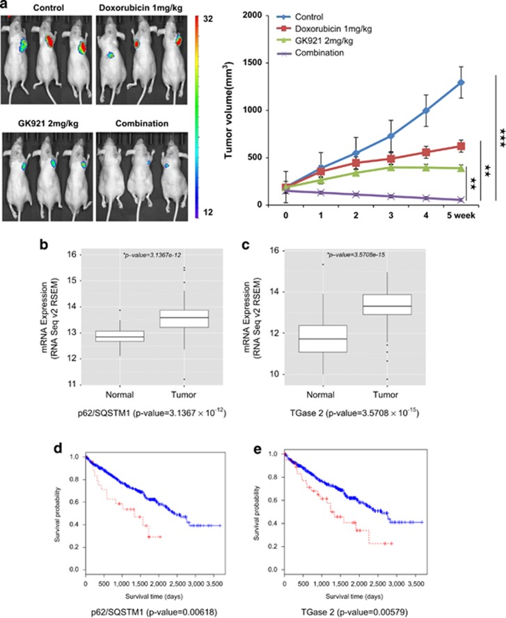
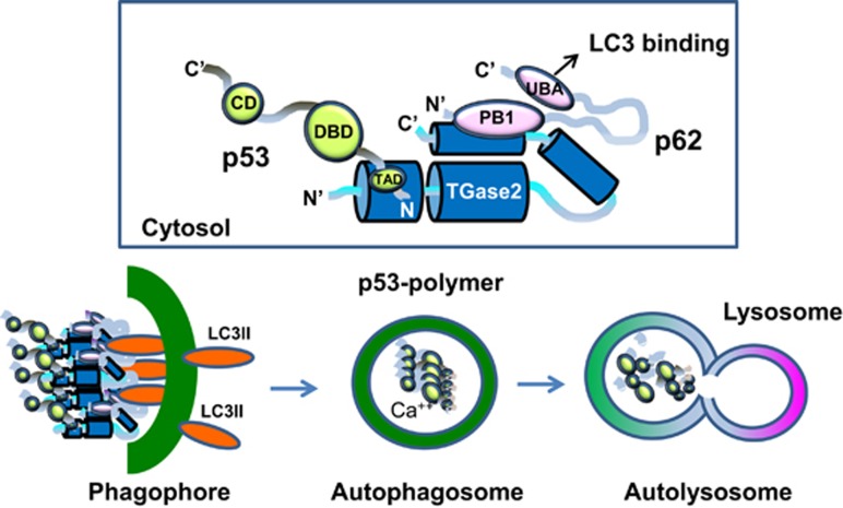
In renal cell carcinoma, transglutaminase 2 (TGase 2) crosslinks p53 in autophagosomes, resulting in p53 depletion and the tumor's evasion of apoptosis. Inhibition of TGase 2 stabilizes p53 and induces tumor cells to enter apoptosis. This study explored the mechanism of TGase 2-dependent p53 degradation. We found that TGase 2 competes with human double minute 2 homolog (HDM2) for binding to p53; promotes autophagy-dependent p53 degradation in renal cell carcinoma (RCC) cell lines under starvation; and binds to p53 and p62 simultaneously without ubiquitin-dependent recognition of p62. The bound complex does not have crosslinking activity. A binding assay using a series of deletion mutants of p62, p53 and TGase 2 revealed that the PB1 (Phox and Bem1p-1) domain of p62 (residues 85-110) directly interacts with the β-barrel domains of TGase 2 (residues 592-687), whereas the HDM2-binding domain (transactivation domain, residues 15-26) of p53 interacts with the N terminus of TGase 2 (residues 1-139). In addition to the increase in p53 stability due to TGase 2 inhibition, the administration of a DNA-damaging anti-cancer drug such as doxorubicin-induced apoptosis in RCC cell lines and synergistically reduced tumor volume in a xenograft model. Combination therapy with a TGase 2 inhibitor and a DNA-damaging agent may represent an effective therapeutic approach for treating RCC.


Figures

Figure 1
Figure 1
TGase 2 and HDM2 regulate p53 stability in an independent manner. ACHN (a and b) and CAKI-1 (c and d) cells were transfected with siRNA targeting TGM2 (a and c) or HDM2 (b and d) for 48 h; then the cells were treated with chloroquine (CQ, 50 μM) or MG132 (10 μM), or left untreated, for 6 h under starvation conditions before harvesting. Whole-cell lysates were subjected to immunoblotting with the indicated antibodies. ACHN (e and f) and CAKI-1 (g and h) cells were treated with siRNA for TGM2 or HDM2, and stained with TUNEL and DAPI (e and g). Scale bar, 20 μm. The percentage of TUNEL-positive cells was counted (f and h). The western blots are representative of three independent experiments. **P<0.01, ***P<0.001
Figure 2
Figure 2
TGase 2 and HDM2 compete for p53 interaction. TGM2 knockdown increased the interaction of p53 with HDM2, whereas it abolished the interaction with p62 (a and b). ACHN and CAKI-1 cells were transfected with siRNA for TGM2 (a) or HDM2 (b) for 48 h under starvation conditions. Whole-cell extracts (left) or p53 immunoprecipitates (right) were subjected to immunoblotting for TGase 2, HDM2, p53 and p62. (c) The induction of DNA damage inhibited the binding of TGase 2 to p53 and induced p53 phosphorylation. CAKI-1 and ACHN cells were treated with doxorubicin (1 μM) or etoposide (50 μM) for 24 h; whole-cell extracts were subjected to p53 immunoprecipitation and western blotting. (d) The activated p53 mimic (S15E) did not interact with TGase 2. CAKI-1 and ACHN were transfected with expression plasmids for p53 (wild type), p53 (S15E), or p53 (S15A) for 24 h, and the cell lysates were immunoprecipitated with a His-tag antibody and subjected to immunoblotting. (e) p53 stabilization (p-p53) by doxorubicin blocked interaction with TGase 2 in a time-dependent manner. After doxorubicin (1 μM) treatment for up to 20 h, cell lysates were immunoprecipitated with an anti-p53 antibody and subjected to immunoblotting. The blots are representative of three independent experiments
Figure 3
Figure 3
TGase 2 chaperones p53 to p62. (a and b) TGase 2 knockdown abolished the interaction of p53 to p62 as well as the interaction of TGase 2 to p53 and p62. TGM2 was silenced in ACHN (a) or CAKI-1 (b) cells for 48 h under starvation conditions, and then cell extracts were subjected to immunoprecipitation of TGase 2, p53, and p62. (c) TGase 2 activity is not required for interacting with p53. Wild-type or catalytically inactive TGase 2 (double mutant, C277S and C370A) was co-transfected with p62 into HEK293 cells. TGase 2 was immunoprecipitated using an anti-HA-tag antibody, followed by immunoblotting of TGase 2, p53 and p62
Figure 4
Figure 4
TGase 2 makes a trimer complex with p53 and p62. (a) The N terminus of p62 (residues 85–110) is the TGase 2-binding site. HEK293 cells were transfected with plasmids encoding HA-tagged TGase 2 (HA-TG2) and either wild-type p62 (p3xFLAG p62 wild type) or a p62 deletion mutant. The proteins were co-immunoprecipitated from cell extracts using an anti-HA-tag antibody and subjected to immunoblotting. (b) The C terminus of TGase 2 is the p62-binding domain (β-barrel domains, residues 461–687). HEK293 cells were transfected with p62 (p3XFLAG p62 wild type) and either wild-type TGase 2 (HA-TG2) or a TGase 2 deletion mutant. The proteins were co-immunoprecipitated from cell extracts using an anti-HA-tag antibody and subjected to immunoblotting. (c) The N terminus of TGase 2 is the p53-binding domain (residues 1–139). HEK293 cells were transfected with wild-type p53 (p3xFLAG p53) and either HA-tagged wild-type TGase 2 (HA-TG2) or a deletion mutant of TGase 2. Proteins were immunoprecipitated using an anti-HA-tag antibody. (d) Primary structures of p62, p53 and TGase 2 proteins. CRD, C-terminal regulatory domain, lysine rich domain; DBD, DNA-binding domain; KIR, Keap1-interacting region; LIR, LC3-interacting region; p53, tumor suppressor; p62, sequestosome 1; TAD, transcription-activation domain or transactivation domain; TBD, TRAF6-binding domain; TET, tetramerization domain; ZZ, zinc-finger domain
Figure 4
Figure 4
TGase 2 makes a trimer complex with p53 and p62. (a) The N terminus of p62 (residues 85–110) is the TGase 2-binding site. HEK293 cells were transfected with plasmids encoding HA-tagged TGase 2 (HA-TG2) and either wild-type p62 (p3xFLAG p62 wild type) or a p62 deletion mutant. The proteins were co-immunoprecipitated from cell extracts using an anti-HA-tag antibody and subjected to immunoblotting. (b) The C terminus of TGase 2 is the p62-binding domain (β-barrel domains, residues 461–687). HEK293 cells were transfected with p62 (p3XFLAG p62 wild type) and either wild-type TGase 2 (HA-TG2) or a TGase 2 deletion mutant. The proteins were co-immunoprecipitated from cell extracts using an anti-HA-tag antibody and subjected to immunoblotting. (c) The N terminus of TGase 2 is the p53-binding domain (residues 1–139). HEK293 cells were transfected with wild-type p53 (p3xFLAG p53) and either HA-tagged wild-type TGase 2 (HA-TG2) or a deletion mutant of TGase 2. Proteins were immunoprecipitated using an anti-HA-tag antibody. (d) Primary structures of p62, p53 and TGase 2 proteins. CRD, C-terminal regulatory domain, lysine rich domain; DBD, DNA-binding domain; KIR, Keap1-interacting region; LIR, LC3-interacting region; p53, tumor suppressor; p62, sequestosome 1; TAD, transcription-activation domain or transactivation domain; TBD, TRAF6-binding domain; TET, tetramerization domain; ZZ, zinc-finger domain
Figure 5
Figure 5
Doxorubicin potentiates the apoptotic effects of TGase 2 silencing by extending p53 stability. (a) CAKI-1 cells were treated with siTGase 2 for 48 h, and then with doxorubicin for an additional 8 h before harvesting for immunoblotting of TGase 2, p53 and downstream markers of p53. (b) Viable cell was counted by using SRB staining after a single treatment of control siRNA, siTGase 2, or doxorubicin (1 μM), or a combined treatment of doxorubicin (1 μM) and siTGase 2. Y-axis presents viable cell per square mm. (c) To measure combination effect of doxorubicin with TGase 2 knockdown against RCC growth, XTT assay was employed under same conditions as (b). (d) To measure induction of apoptosis by dual treatments, CAKI-1 and ACHN cells were treated with a combination of GK921 and doxorubicin (1 μM) for 0, 8, 12, 18 and 20 h. Immunoblotting was performed against cleaved PARP and p-p53. (e) To measure combination effect of doxorubicin and GK921 against RCC growth, XTT assay was employed under same conditions as (d). Cumulative data from three independent experiments is shown here as mean±S.D. (n=3). *P<0.05, **P<0.01, ***P<0.001
Figure 6
Figure 6
Doxorubicin amplifies the therapeutic response of RCC to the TGase 2 inhibitor GK921. (a) ACHN-luciferase cells were injected subcutaneously into 6–8-week-old female BALB/c nude mice. When the tumor volume reached 100 mm3, mice were treated orally, 5 days/week, with GK921 (2 mg/kg/day), doxorubicin (1 mg/kg/day), both or vehicle (n=3 per each group). After 5 weeks of treatment, tumor size was measured by the photon flux from luciferin and expressed on a color scale and as photons/s/cm2/steradian (left). Tumor volume was also measured using calipers (right). (be) Bioinformatics analysis of p62 gene (SQSTM1) and TGase 2 gene (TGM2) expression in clear cell RCC patients. (b) Lower p62/SQSTM1 expression in normal kidney samples than in matched clear cell RCC samples (P=3.1367 × 10⁻¹²). (c) Lower TGM2 expression in normal kidney samples than in matched clear cell RCC samples (P=3.5708 × 10⁻¹⁵). (d) Patients with high levels of p62/SQSTM1 expression (red) had lower overall survival (OS) than those with low levels (blue). Log-rank test, P=0.00618. (e) Patients with high levels of expression of TGM2 (red) also had a reduced OS. Gene expressions are colored red; higher levels of gene expression cut off z-score>2σ. Log-rank test, P=0.00579
Figure 7
Figure 7
Model of the interactions of TGase 2, p53 and p62 based on the experimental data. The N terminus of TGase 2 interacts with the N terminus of p53, and the C terminus of TGase 2 interacts with the N terminus of p62 simultaneously. The C terminus of p62 in the complex is free and transfers the complex to LC3 in the phagophore. P53 is polymerized in the autophagosome when calcium increases. Later, the autophagosome and lysosome fuse into an autolysosome, which degrades all crosslinked materials. The TGase 2-binding site of p53 was identified as the HDM2-binding site in Figures 2d and e. Previously, we found that the TGase 2-targeting site is the DBD domain. CD, complexing domain; DBD, DNA-binding domain

References

    1. Folk JE. Transglutaminases. Annu Rev Biochem 1980; 49: 517–531. - PubMed
    1. Ku BM, Kim DS, Kim KH, Yoo BC, Kim SH, Gong YD et al. Transglutaminase 2 inhibition found to induce p53 mediated apoptosis in renal cell carcinoma. FASEB J 2013; 27: 3487–3495. - PubMed
    1. Ku BM, Kim SJ, Kim N, Hong D, Choi YB, Lee SH et al. Transglutaminase 2 inhibitor abrogates renal cell carcinoma in xenograft models. J Cancer Res Clin Oncol 2014; 140: 757–767. - PMC - PubMed
    1. Oliner JD, Pietenpol JA, Thiagalingam S, Gyuris J, Kinzler KW, Vogelstein B. Oncoprotein MDM2 conceals the activation domain of tumour suppressor p53. Nature 1993; 362: 857–860. - PubMed
    1. Haupt Y, Maya R, Kazaz A, Oren M. Mdm2 promotes the rapid degradation of p53. Nature 1997; 387: 296–299. - PubMed

Publication types

MeSH terms