. 2016 Mar 31;11(3):e0152967.
doi: 10.1371/journal.pone.0152967. eCollection 2016.

An Ultrastructural and Immunohistochemical Analysis of the Outer Plexiform Layer of the Retina of the European Silver Eel (Anguilla anguilla L)

Affiliations

An Ultrastructural and Immunohistochemical Analysis of the Outer Plexiform Layer of the Retina of the European Silver Eel (Anguilla anguilla L)

Jan Klooster et al. PLoS One. .

Abstract

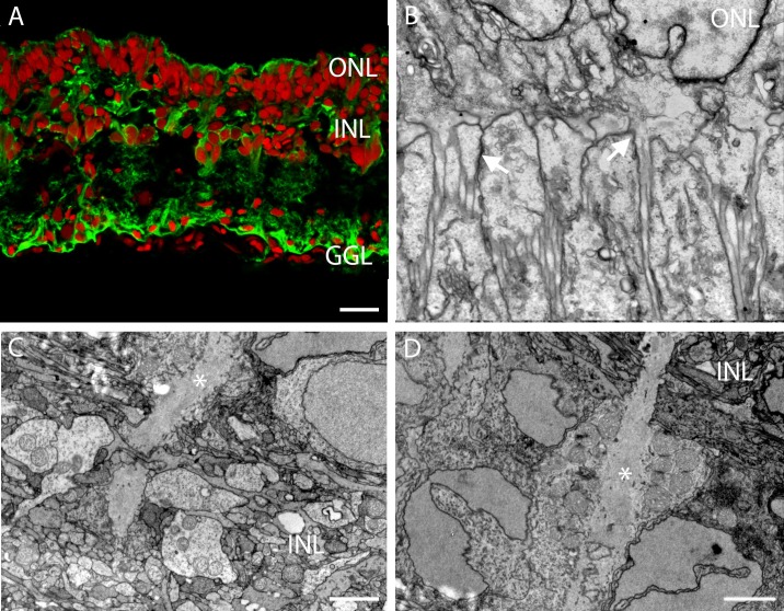
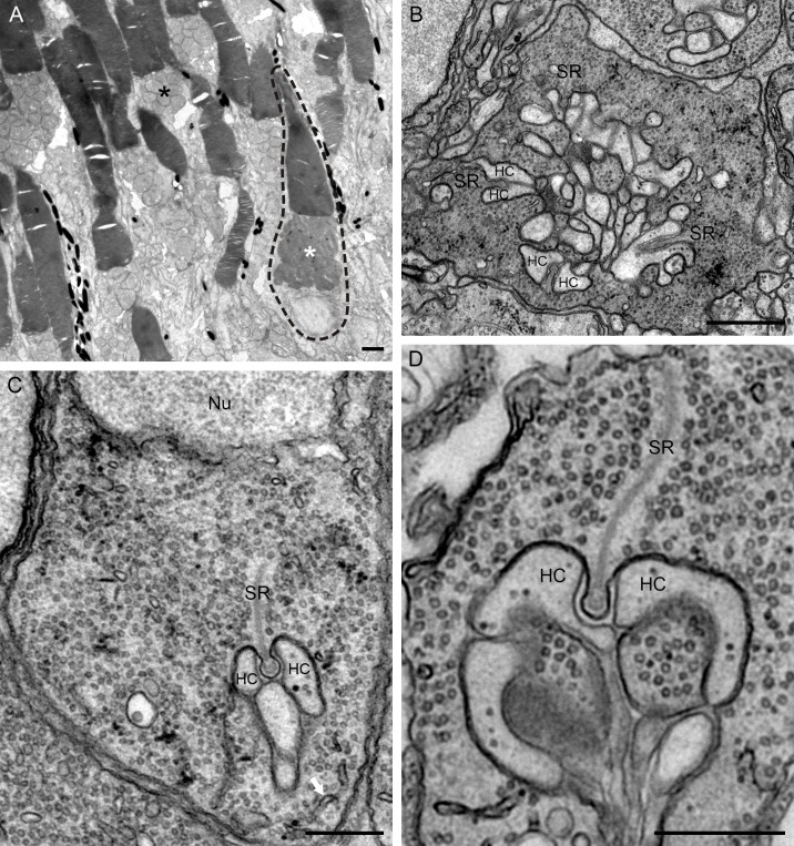
Here we studied the ultrastructural organization of the outer retina of the European silver eel, a highly valued commercial fish species. The retina of the European eel has an organization very similar to most vertebrates. It contains both rod and cone photoreceptors. Rods are abundantly present and immunoreactive for rhodopsin. Cones are sparsely present and only show immunoreactivity for M-opsin and not for L-, S- or UV-cone opsins. As in all other vertebrate retinas, Müller cells span the width of the retina. OFF-bipolar cells express the ionotropic glutamate receptor GluR4 and ON-bipolar cells, as identified by their PKCα immunoreactivity, express the metabotropic receptor mGluR6. Both the ON- and the OFF-bipolar cell dendrites innervate the cone pedicle and rod spherule. Horizontal cells are surrounded by punctate Cx53.8 immunoreactivity indicating that the horizontal cells are strongly electrically coupled by gap-junctions. Connexin-hemichannels were found at the tips of the horizontal cell dendrites invaginating the photoreceptor synapse. Such hemichannels are implicated in the feedback pathway from horizontal cells to cones. Finally, horizontal cells are surrounded by tyrosine hydroxylase immunoreactivity, illustrating a strong dopaminergic input from interplexiform cells.


Conflict of interest statement

Competing Interests: The authors have declared that no competing interests exist.

Figures

Fig 1
Fig 1. Lightmicrograph of a 1 μm thick section of the posterior eye segment of the European eel.
The tissue is embedded in epoxy resin, stained with toluidine blue. From top to bottom one can identify the sclera, the outer segments of the photoreceptors (OS), the inner segments of the cones (arrows), the outer nuclear layer (ONL), the outer plexiform layer (OPL), the inner nuclear layer (ONL), the inner plexiform layer (IPL and the ganglion cell layer (GCL). Note the cartilage in the sclera (white asterisk). Scale bar represents 17 μm.
Fig 2
Fig 2. Abundant presence of Mΰller cells in the retina of the European silver eel.
A) Confocal picture of the glutamine synthetase immunoreactivity (green). The red label is a nuclear stain. All layers of the retina show thick glutamine synthetase immunoreactive processes. B) Electronmicrograph of the retinal ONL. Junction are made by Mΰller cell processes and photoreceptor processes (white arrows). C) Electronmicrograph showing a Mΰller cell (white asterisk) containing mitochondria at the level of the IPL. D) Electronmicrograph showing a Mΰller cell process (white asterisk) at the level of the INL. Note that this process contains many mitochondria. Scale bar in Fig 2A represents 20 μm, scale bar in Fig 2B represents 1 μm, and scale bars in Fig 2C and 2D represent 2 μm.
Fig 3
Fig 3. Confocal pictures of the retina of the European eel stained with the M-opsin and the rhodopsin antibodies.
A) M-opsin labeling of cone outer segments in a section of the retina (M-opsin: green; Nuclei: red). B) Combined picture of lightmicroscopical image and fluorescence image showing that only cones have M-opsin immunoreactivity. C) M-opsin labeling (green) in a flat mounted section of peripheral retina. D) Rod rhodopsin labeling (green; nuclei: red) is abundantly present in the retina of the European eel. Scale bars in A, C and D represent 5 μm and in B represents 6 μm.
Fig 4
Fig 4. Electronmicrographs of the retina of the European eel.
A) The outer and inner segments of the photoreceptors. A cone is encircled with a dotted line while the remained are rods. Note that the mitochondria in the cone inner segments are electron dense (white asterisk) whereas they are electron lucent in rod inner segments (black asterisk). B) Cone pedicle with several ribbon synapses (SR). C) and D) rod spherules (Nu: Nucleus). The white arrow in C (lower right corner) points to the smooth endoplasmic reticulum. The lateral element of the synaptic triad representing horizontal cell dendrites (HC) can transfers to central element. Bars in panels A and B represent 1 μm, bars in panels C and D represent 0.5 μm.
Fig 5
Fig 5. Combined panel of confocal pictures and electronmicrographs focused on horizontal cells.
A) Immunofluorescence of the calretinin antibody (green; red: nuclei). Strong labeling occurred in the IPL and some amacrine cells were also labeled. Note, however, the labelling in the horizontal somata and the dendrites (white asterisks). B) Horizontal cells (red: nuclei; white asterisks) are surrounded by TH-immunoreactive processes (green). Additionally, the TH-immunoreactivity in the IPL is indicative of interplexiform cells. C) Punctate labeling of Cx53.8 (green) around the somata of horizontal cells at the level of the OPL. D) and E) Electronmicrographs of the Cx53.8 immunoreactivity. D) Lower magnification of the neuropil of the OPL of the European silver eel retina, showing many Cx53.8 immunoreactive gap junctions. E) Higher magnification of a gap junction. Note that the membranes are separated at the borders of the gap-junction and then come very close together at position of the gap junction itself. At that location the labeling is the strongest. Scale bars in panels A and B represent 5 μm, scale bar in panel C represents 10 μm and scale bars in panel D and E represent 0.25 μm.
Fig 6
Fig 6. Electronmicrographs of the CX53.8 immunoreactivity at the cone pedicle and the rod spherule level.
A) Cone pedicle with Cx53.8 immunoreactivity at one laterally ending horizontal cell dendrite. Note that the opposing horizontal cell dendrite is not labeled. B) and C) Rod spherules with Cx53.8 immunoreactivity in the laterally ending horizontal cell dendrites. In rods, both lateral elements show Cx53.8 immunoreactivity. Synaptic ribbon (SR); horizontal cell (HC). Scale bars represent 0.25 μm.
Fig 7
Fig 7. Combined panel of confocal picture and electronmicrographs of the GluR4 immunoreactivity.
A) Confocal picture of GluR4 immunoreactivity (green; nuclei: red) in the OPL of the European silver eel retina. Note the punctate labeling of the GluR4 immunoreactivity (white arrows). Mϋller cells also show GluR4 immunoreactivity (white arrowheads). B) Electronmicrograph of a cone pedicle; GluR4 immunoreactivity is seen at the base of the cone pedicle whereas the lateral elements were not labeled. C) Electronmicrograph of a rod spherule. GluR4 immunoreactivity was found in the central element representing the OFF-bipolar cell dendrite. Synaptic ribbon (SR); horizontal cell (HC). Scale bar in panel A represents 5 μm, and scale bars in panel B and C represent 0.25 μm.
Fig 8
Fig 8. Confocal picture and electronmicrographs of the PKCα immunoreactivity.
A) Confocal picture of PKCα immunoreactivity (green; red: nuclei). Note that the entirety of the ON-bipolar cells are labeled from the dendrites in the OPL via the cell somata in INL to the axon terminals in the IPL. Two types of axon terminal show PKCα immunoreactivity, one type is more bulbous (small white asterisks) while the other is smaller (white arrowhead). B) Electronmicrograph of a cone pedicle with PKCα immunoreactivity at the position of the central elements representing ON-bipolar cell dendrites. C) and D) Electronmicrographs of rod spherules with PKCα immunoreactivity at the position of the central elements representing the ON-bipolar cell dendrite. No labeling was found in the lateral elements. Synaptic ribbon (SR); horizontal cell (HC). Scale bar in panel A represents 5 μm, and scale bars in panels B, C and D represent 0.25 μm.
Fig 9
Fig 9. Panel of a confocal picture and electronmicrographs of the mGluR6 immunoreactivity.
A) Confocal picture of the mGluR6 immunoreactivity (green; red: nuclei) showing punctate labeling the OPL and two diffuse bands in the IPL. B) Electronmicrograph of a cone pedicle. mGluR6 immunoreactive processes were observed at the base of the cone pedicle. No mGluR6 immunoreactivity was found at the lateral elements representing the horizontal cell dendrites. C) and D) Electronmicrographs of rod spherules showing the mGluR6 immunoreactivity on the central elements of the triad representing the bipolar cell dendrites. Synaptic ribbon (SR); Horizontal cell (HC); Nucleus (Nu). Scale bar in panel A represents 5 μm, bars in panels B, C and D represent 0.25 μm.
Fig 10
Fig 10. Confocal pictures of double label experiments with the antibodies against mGluR6 and PKCα.
A) Retinal section double labeled with antibodies against mGluR6 (red) and PKCα (green) shows co-localization. B) mGluR6 immunoreactivity (red). C) PKCα immunoreactivity (green). Note the clear punctate co-localization in the OPL. No co-localization in the IPL. Scale bar represents 5 μm.

Similar articles

Cited by

References

    1. Aoyama J (2009) Life history and evolution of migration in catadromous eels (Genus Anguilla). Aqua-BioSci Monogr 2: 1–42.
    1. Van Ginneken VJT, Maes GE (2005) The European eel (Anguilla anguilla, Linaeus), its lifecycle, evolution and reproduction: A literature review. Rev in Fish Biol & Fisheries 15: 367–398.
    1. Tesch F- W (1977) Body structure and functions In: Greenwood PH, editor. The Eel Biology and management of anguillid eels. London: Chapman; pp. 68–73.
    1. Rousseau K, Aroua S, Schmitz M, Elie P, Dufour S (2012) Silvering metamorphosis or puberty In: dvan den thillart G, Dufour S, Rankin JC, editors. Spawning mirgation of the european eel. Netherlands: Spinger; pp. 39–63.
    1. Endeman D, Klaassen LJ, Kamermans M (2013) Action spectra of zebrafish cone photoreceptors. PLoS One 8: e68540 10.1371/journal.pone.0068540 - DOI - PMC - PubMed

MeSH terms