. 2016 Apr 1:6:23603.
doi: 10.1038/srep23603.

DNA damage regulation and its role in drug-related phenotypes in the malaria parasites

Affiliations

DNA damage regulation and its role in drug-related phenotypes in the malaria parasites

Devendra Kumar Gupta et al. Sci Rep. .

Abstract

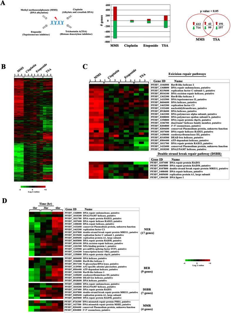
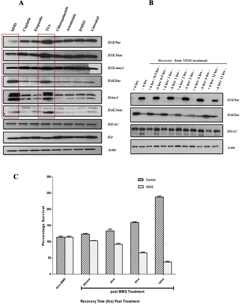
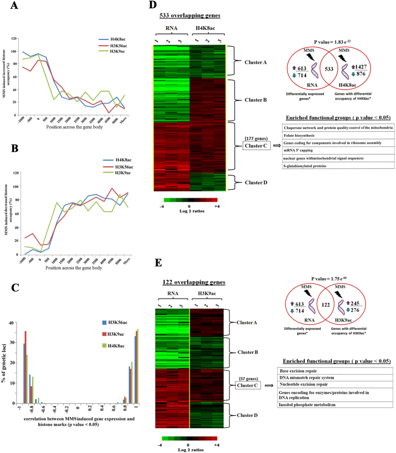
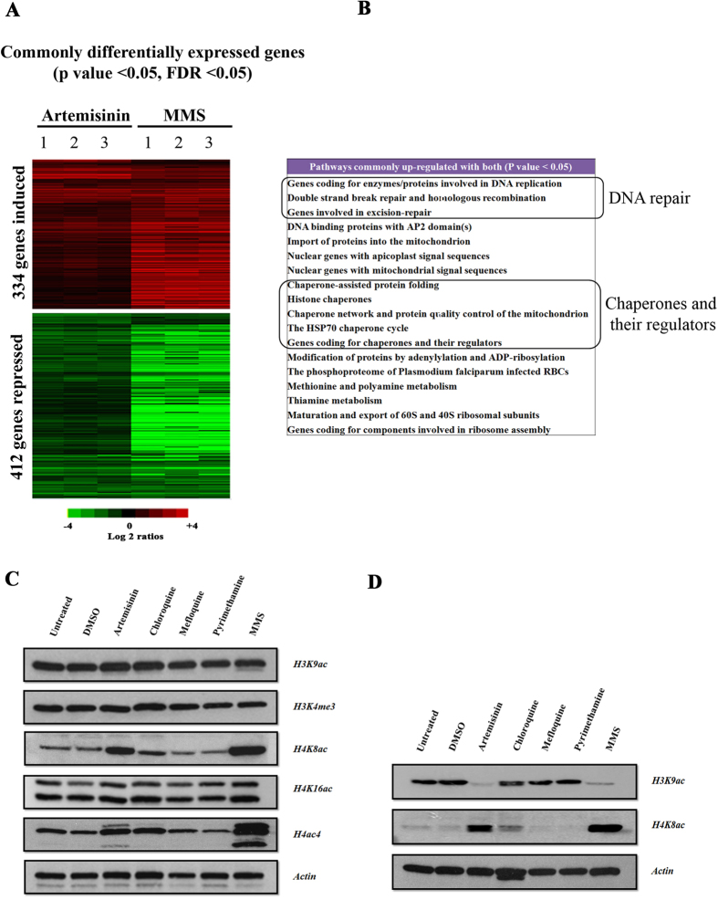
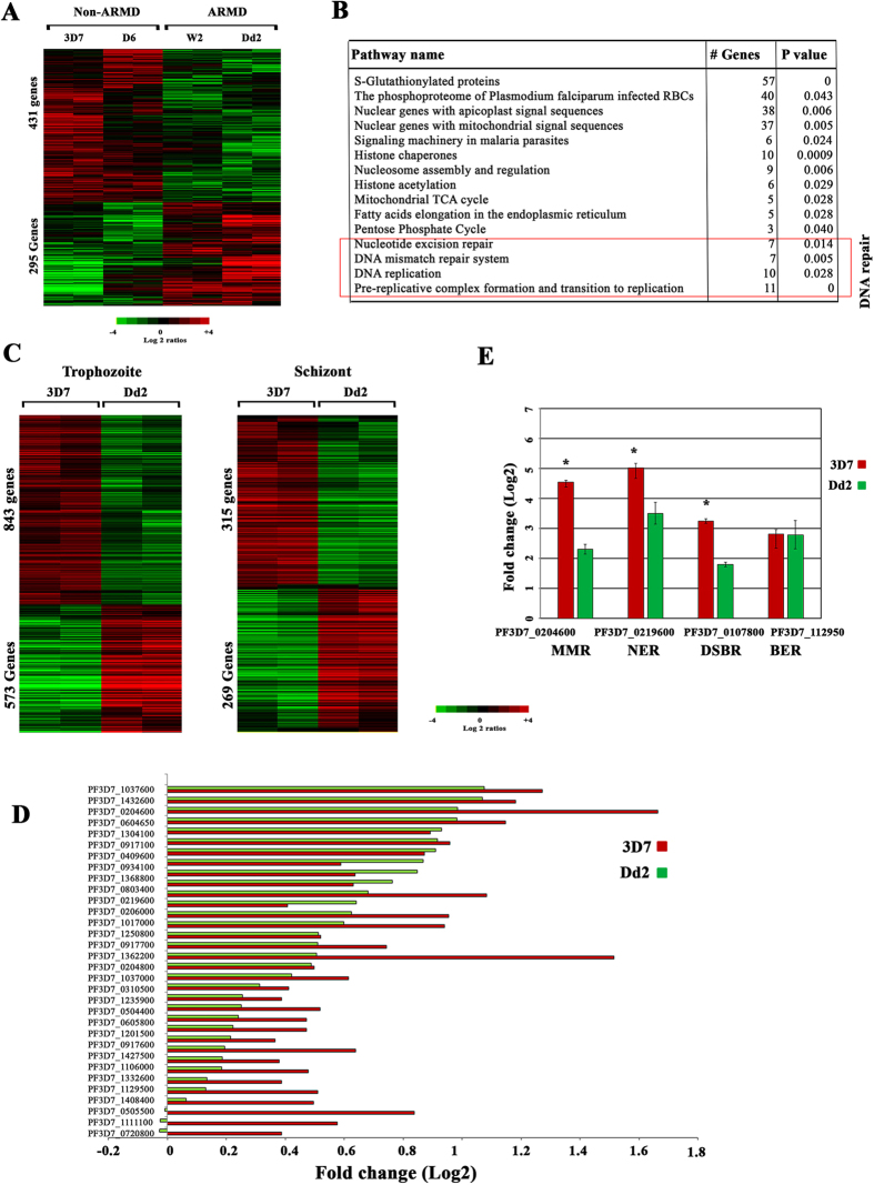
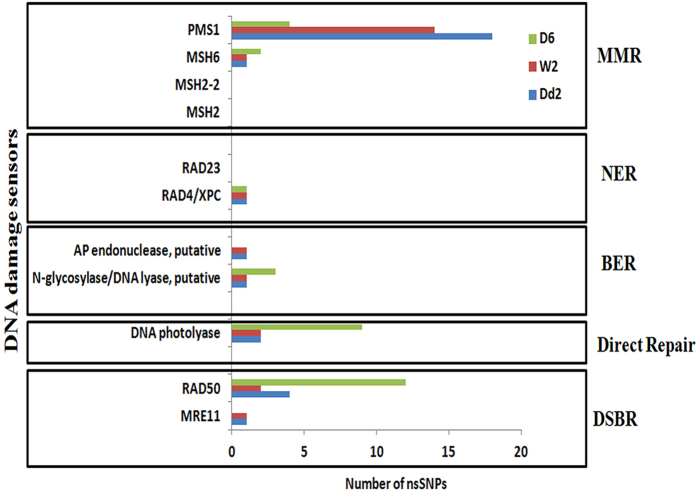
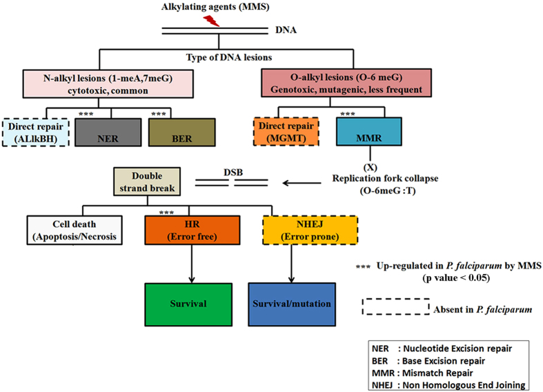
DNA of malaria parasites, Plasmodium falciparum, is subjected to extraordinary high levels of genotoxic insults during its complex life cycle within both the mosquito and human host. Accordingly, most of the components of DNA repair machinery are conserved in the parasite genome. Here, we investigated the genome-wide responses of P. falciparum to DNA damaging agents and provided transcriptional evidence of the existence of the double strand break and excision repair system. We also showed that acetylation at H3K9, H4K8, and H3K56 play a role in the direct and indirect response to DNA damage induced by an alkylating agent, methyl methanesulphonate (MMS). Artemisinin, the first line antimalarial chemotherapeutics elicits a similar response compared to MMS which suggests its activity as a DNA damaging agent. Moreover, in contrast to the wild-type P. falciparum, two strains (Dd2 and W2) previously shown to exhibit a mutator phenotype, fail to induce their DNA repair upon MMS-induced DNA damage. Genome sequencing of the two mutator strains identified point mutations in 18 DNA repair genes which may contribute to this phenomenon.


Figures

Figure 1
Figure 1. Global transcriptional response of P. falciparum to DNA damaging agents.
(A) Four DNA and/or chromatin perturbation agents MMS (0.05%), Cisplatin (100 μM), Etoposide (100 μM), and TSA (50 μM) were chosen to study the DNA damage response of P. falciparum parasites. The bar graph represents the number of genes differentially induced by >2 fold after synchronized parasites were treated at the trophozoite stage for 6 hr. The Venn diagram represents differentially expressed genes between the MMS or TSA treatments. (B) Heat map represents up (red) and down (green) regulated genes induced by the drug treatments by >2-fold. The results are mean value of a relative mRNA abundance in three experimental replicates. (C) The expression pattern of 29 excision and 6 DSB repair genes which were up-regulated by MMS are shown along with the effect of cisplatin, etoposide and TSA among three independent replicates (p-value < 0.05). (D) The heat map represents differential expression of DNA repair genes in a time course experiment with samples collected at 1, 3 and 6 hr of the MMS (0.05%) treatment. Only with the 6 hr of exposure to MMS, DNA repair machinery was up-regulated with 9 BER, 17 NER, 4 MMR and 7 DBSR genes significantly up-regulated in the two independent replicates (p- value 0.0002–0.02).
Figure 2
Figure 2. DNA damage affects the overall abundance of histone modifications during the P. falciparum IDC.
(A) 3D7 parasites were treated with all drugs at the trophozoite stage for 6 hr and the overall abundance of individual histone modification was measured by western blotting. The MMS treatment resulted in reduction of H3K9ac and H3K56ac and increase of H4K8ac, H4K16ac and H4ac4. Conversely, TSA induced hyperacetylation of all studied H3 and H4 acetylations. Cisplatin and etoposide as well as artemisinin and chloroquine at IC-90 concentrations had no effect on the overall abundance on the histone modification compared to P. falciparum grown under normal condition or treated with the carrier solvent, DMSO. Specific antibody for HDAC enzyme, histone 4 and actin were used as loading controls. (B) Recovery dynamics of H3K9ac and H4K8ac were assessed by first treating with MMS for 6 hrs and subsequently drug was removed and samples were collected after 0.5 hrs, 1 hrs, 2 hrs, 6 hrs and 12 hrs. (C) Parasite survival after MMS (0.05%) treatment compared to untreated 3D7 (Control) parasites. Graphical representation shown is percentage survival in MMS treated and control parasites at 6hrs MMS and 30 mins, 2 hrs, 6 hrs and 12 hrs (post-MMS wash off treatment). The error bars represent SEM.
Figure 3
Figure 3. DNA damage induces genome wide spread of H4K8 acetylation whereas H3K9 acetylation preferentially marks the DNA repair genes.
(A,B) The horizontal axis is the microarray oligonucleotide probes (MOE) position relative to the ATG start codon. The vertical axis is the number of histone marks associated genetic loci normalized to the number of probes which represent that particular region on the genome and these genetic loci are those which have increased (A) or decreased (B) ChIP signal in presence of MMS (p value < 0.05). (C) The correlation between MMS induced gene expression and histone marks is shown. (D,E) Association between differential expression and altered histone marks occurrence during MMS treatment is shown. To study such association, we correlated ChIP-on-chip dataset and RNA expression dataset obtained from MMS treated trophozoite parasites and it results in significant overlap between up-regulated/down-regulated genes (>2 fold, p value < 0.05) due to MMS treatment and with those that had altered histone marks (p value < 0.05). For H4K8ac (D) and H3K9ac (E), we identified 533 and 122 overlapping genes. Hierarchical clustering and enriched functional groups of these 533 and 122 genes were also shown, which are divided into 4 clusters depending upon on correlation between ChIP-on-chip and transcriptome dataset.
Figure 4
Figure 4. Artemisinin induces DNA damage response in P. falciparum.
(A) The synchronized P. falciparum cells were treated with clinical doses of artemisinin (1 μM) and MMS (0.05%) for 6 hr at the trophozoite stage. Genes with similar differential transcription for both MMS and artemisinin are shown in the heat map. The experiments were done in triplicates. (p value < 0.05, FDR < 0.05) (B) Pathways, which are commonly up-regulated by both artemisinin and MMS. Schizont (C) and Trophozoite stages (D) of parasites were treated with clinical doses of different anti-malarial drug for 6 hr: artemisinin (1 μM), chloroquine (30 μM), mefloquine (5 μM) and pyrimethamine (5 μM) and with MMS (0.05%). Immunoblot analyses were carried out using primary antibodies probed against the core histone modifications.
Figure 5
Figure 5. ARMD parasites displaying mutator phenotype have defective DNA repair.
(A) P. falciparum ARMD and non-ARMD parasites were treated with MMS (0.05%) at trophozoite stage for 6 hr. Heat map showing the MMS induced differentially expressed genes between the ARMD and non-ARMD parasites (p value < 0.05) at trophozoite stage. Shown are the mean-centered expression log2 ratios for these genes. (B) Functional enrichment analysis on the 431 up-regulated genes is shown. (C) The gene expression patterns of differentially expressed genes at trophozoite and schizont stage are shown between 3D7 and Dd2. (D) 34 repair genes, which were differentially expressed, were plotted for ARMD (Dd2) and non-ARMD (3D7). (E) Validation of differential expression of genes between 3D7 and Dd2 was done by Quantitative RT-PCR. Quantitative RT-PCR was done on four genes using RNA derived from samples treated with MMS for 6 hr. PF3D7_0204600 belongs to mismatch repair (MMR), PF3D7_0219600 belong to nucleotide excision repair (NER), PF3D7_0107800 belong to double strand break repair (DSBR) and PF3D7_112950 belongs to base excision repair (BER). Relative fold change in RNA expression was computed by 2−∆∆Ct method. The reference control gene used was PF3D7_1218600 (arginyl-tRNA synthetase). Error bars shows the standard deviation of the 2−∆∆Ct value over triplicates. (*represents genes which were differentially expressed between 3D7 and Dd2).
Figure 6
Figure 6. DNA polymorphism in DNA damage sensors between ARMD and non-ARMD parasites.
There are three different repair mechanisms: DNA excision repair (BER, MMR, NER), double strand break repair (DSBR) and direct repair (DNA photolyase). Each repair mechanism has different set of sensory proteins which detect DNA damage, for example RAD23 and XPC are the DNA damage recognition proteins for NER repair mechanism. Each box have the distribution of SNP in DNA damage sensory proteins for a specific repair pathway among D6, 3D7 (non-ARMD) and Dd2, W2 (ARMD). Also, included amino acid mutation which changed form reference.
Figure 7
Figure 7. Overview of the P. falciparum DNA damage pathways and their transcriptional properties under MMS-induced stress.
The initial N- and O-alkyl lesions of P. falciparum DNA caused by MMS leads initially to transcriptional induction of nucleotide excision repair (NER), base excision repair (BER) and mismatch repair (MMR). The subsequent induction of the double strand break repair (DSBR), namely the homologous recombination repair (HR), suggests that the MMS induced O-alkyl lesions was not be fully repaired by MMR. The transcriptional induction encompasses the majority (if not all) components of the P. falciparum DNA damage repair. Other components of eukaryotic DNA repair pathways such as nonhomologous end joining (NHEJ), or enzymes of direct repair (ALlkBH and MGMT) are not present in the P. falciparum genome (dashed line outline). Although extensive DSB could lead to cell death, 6 hr treatment with 0.05% MMS was likely counteracted by the DNA repair factors and did not lead to upregulation of putative factors of P. falciparum cell death. ***indicates statistically significant functional enrichment of genes of particular pathway amongst genes upregulated as a result of MMS-induced DNA damage. This outline is based on bioinformatics sequence analysis and the transcriptional results presented in this study.

References

    1. Nowsheen S. & Yang E. S. The Intersection between DNA damage response and cell death pathways. Exp Oncol 34, 243–254 (2012). - PMC - PubMed
    1. Ataian Y. & Krebs J. E. Five repair pathways in one context: chromatin modification during DNA repairThis paper is one of a selection of papers published in this Special Issue, entitled 27th International West Coast Chromatin and Chromosome Conference, and has undergone the Journal’s usual peer review process. Biochem Cell Biol 84, 490–494 (2006). - PubMed
    1. Lee A. H., Symington L. S. & Fidock D. A. DNA Repair Mechanisms and Their Biological Roles in the Malaria Parasite Plasmodium falciparum. Microbiol Mol Biol Rev 78, 469–486 (2014). - PMC - PubMed
    1. Percário S. et al. Oxidative Stress in Malaria. Int J Mol Sci 13, 16346–16372 (2012). - PMC - PubMed
    1. Radfar A., Diez A. & Bautista J. M. Chloroquine mediates specific proteome oxidative damage across the erythrocytic cycle of resistant Plasmodium falciparum. Free Radic Biol Med 44, 2034–2042 (2008). - PubMed

MeSH terms