. 2016 Apr 1:7:11091.
doi: 10.1038/ncomms11091.

p53 downregulates the Fanconi anaemia DNA repair pathway

Affiliations

p53 downregulates the Fanconi anaemia DNA repair pathway

Sara Jaber et al. Nat Commun. .

Abstract

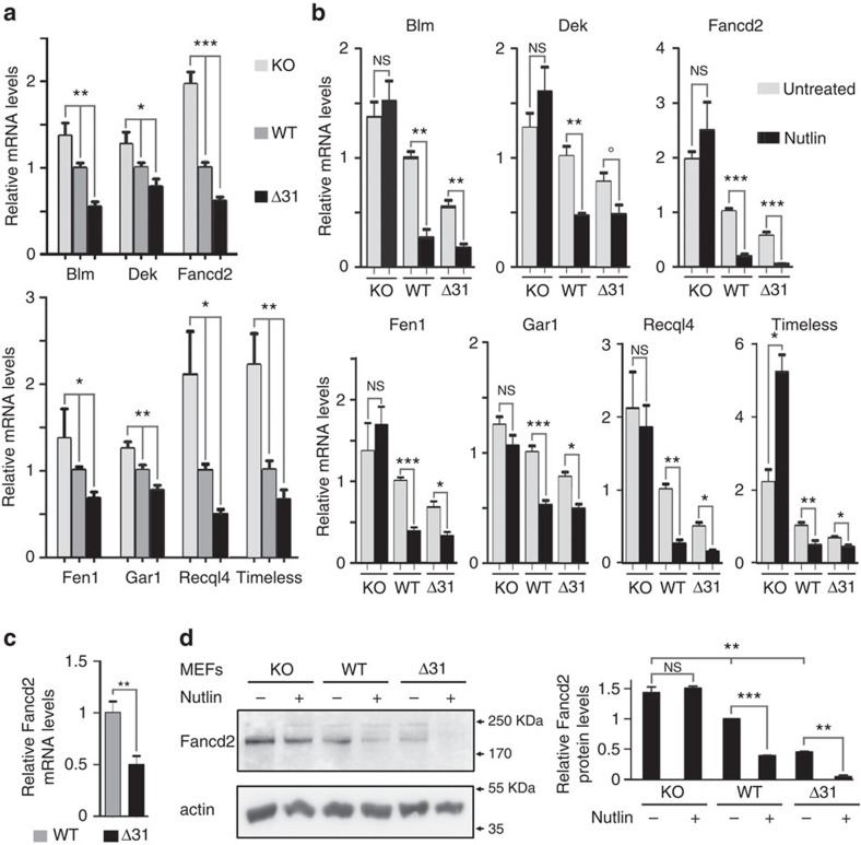
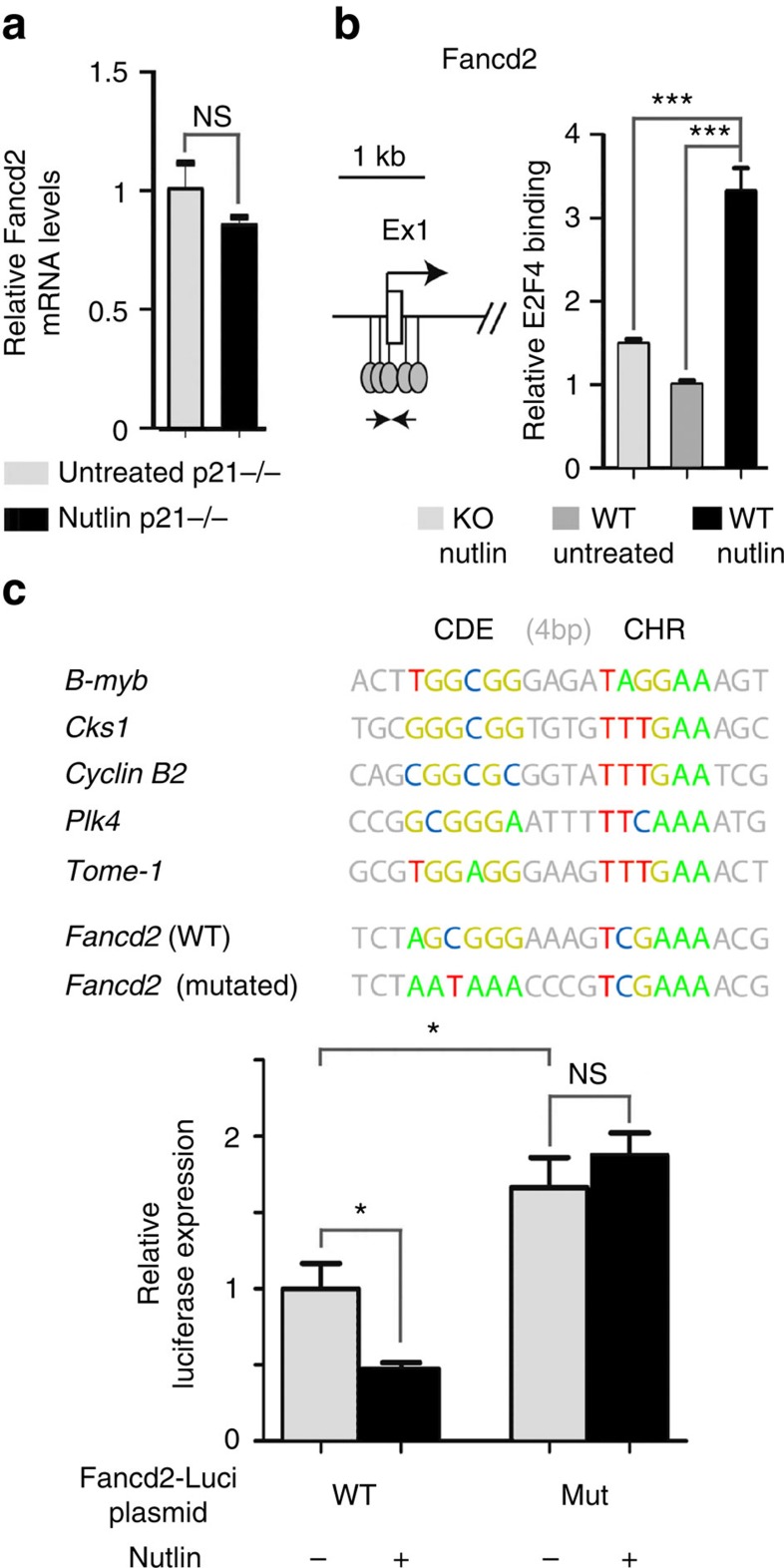
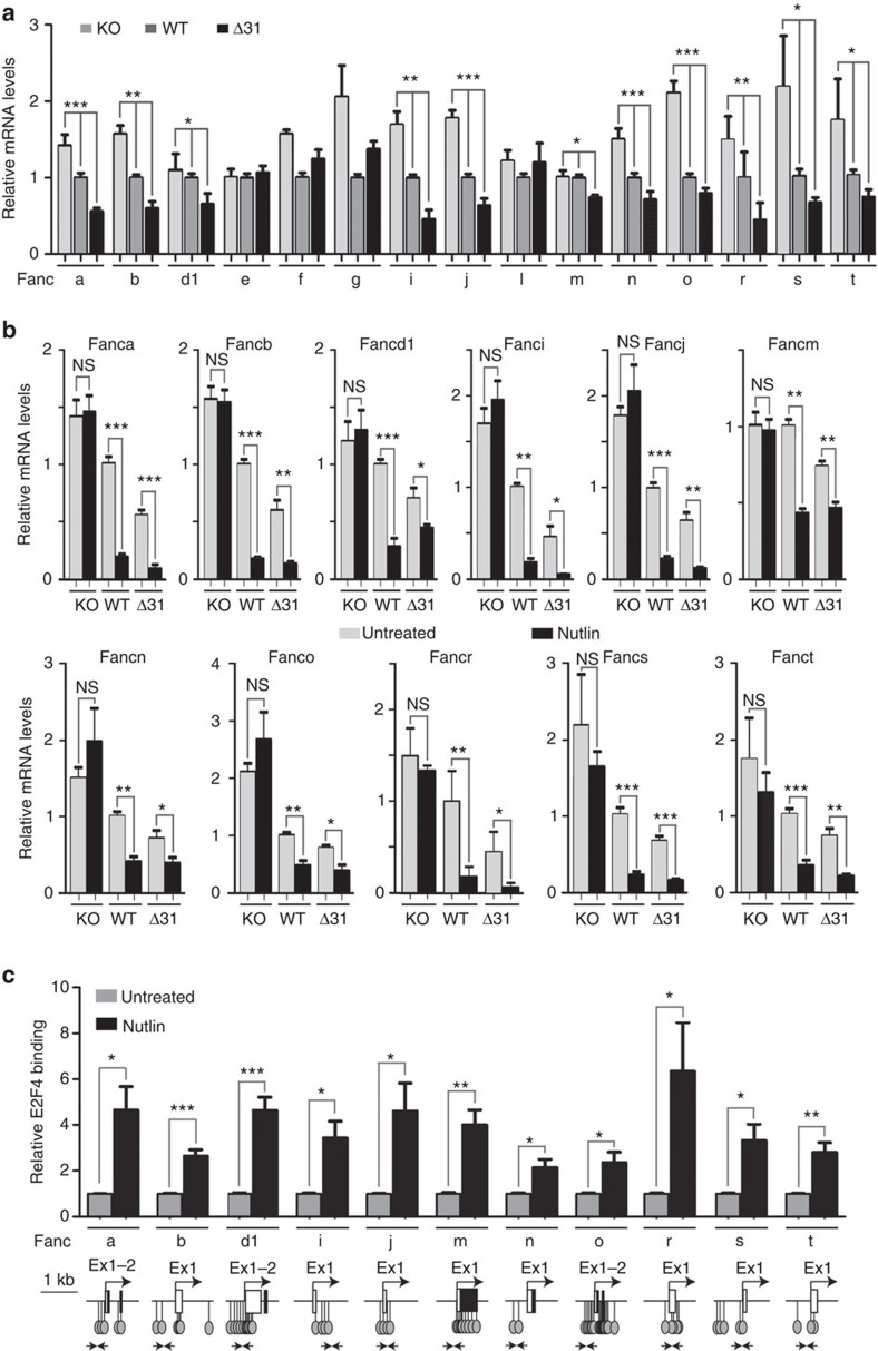
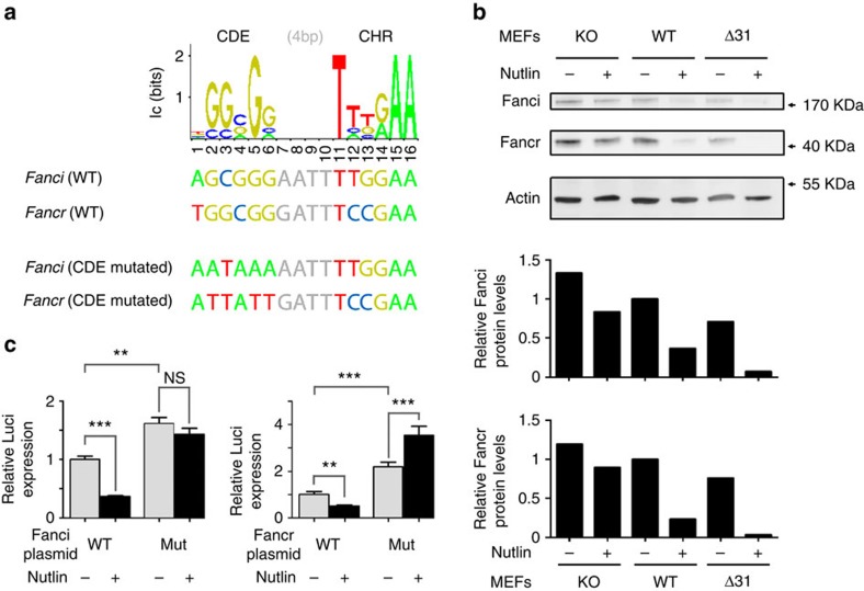
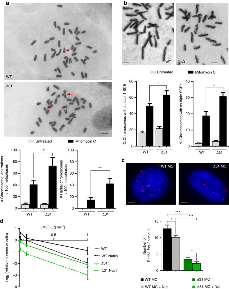
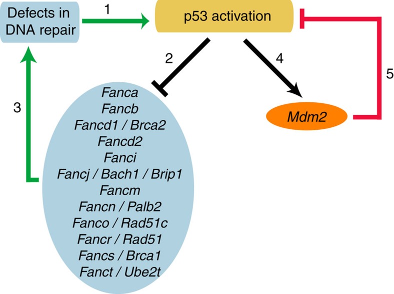
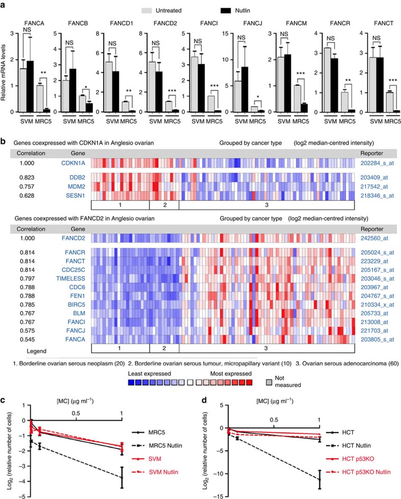
Germline mutations affecting telomere maintenance or DNA repair may, respectively, cause dyskeratosis congenita or Fanconi anaemia, two clinically related bone marrow failure syndromes. Mice expressing p53(Δ31), a mutant p53 lacking the C terminus, model dyskeratosis congenita. Accordingly, the increased p53 activity in p53(Δ31/Δ31) fibroblasts correlated with a decreased expression of 4 genes implicated in telomere syndromes. Here we show that these cells exhibit decreased mRNA levels for additional genes contributing to telomere metabolism, but also, surprisingly, for 12 genes mutated in Fanconi anaemia. Furthermore, p53(Δ31/Δ31) fibroblasts exhibit a reduced capacity to repair DNA interstrand crosslinks, a typical feature of Fanconi anaemia cells. Importantly, the p53-dependent downregulation of Fanc genes is largely conserved in human cells. Defective DNA repair is known to activate p53, but our results indicate that, conversely, an increased p53 activity may attenuate the Fanconi anaemia DNA repair pathway, defining a positive regulatory feedback loop.


Figures

Figure 1
Figure 1. p53 activation leads to the downregulation of seven telomere-related genes.
(a) A comparison of p53−/−, wild-type and p53Δ31/Δ31 cells suggests the p53-dependent regulation of Blm, Dek, Fancd2, Fen1, Gar1, Recql4 and Timeless. RNAs, prepared from unstressed p53−/− (KO), wild-type (WT) and p53Δ31/Δ31 (Δ31) MEFs, were used to compare the expression of 42 genes with a proposed impact on telomere metabolism. mRNAs were quantified using real-time PCR, normalized to control mRNAs, then the amount in WT cells was assigned a value of 1. Shown here are the seven genes for which the mean mRNA levels were intermediate in WT cells compared with p53−/− and p53Δ31/Δ31 cells, with significant differences between the means according to one-way analysis of variance (ANOVA). For the 35 genes that did not match these criteria, see Supplementary Fig. 1. Results from ⩾3 independent experiments. (b) The seven genes are downregulated upon p53 activation. mRNAs were quantified in p53−/−, WT and p53Δ31/Δ31 MEFs, untreated or treated with 10 μM Nutlin for 24 h. Results from ⩾3 independent experiments. °P=0.059. (c) Fancd2 mRNAs are decreased in the bone marrow cells (BMCs) of p53Δ31/Δ31 mice. Fancd2 mRNAs were quantified from the BMCs of nine WT and six p53Δ31/Δ31 mice. (d) p53 activation leads to decreased Fancd2 protein levels. Protein extracts, prepared from untreated or Nutlin-treated MEFs, were immunoblotted with antibodies against Fancd2 and actin. On the left, a typical western blot is shown; on the right, bands from two western blots were quantified and the amount of Fancd2 in unstressed WT cells was assigned a value of 1. In all figures, means+s.e.m. are shown; ***P≤0.001, **P≤0.01, *P≤0.05, NS, not significant by analysis of variance or Student's t-tests.
Figure 2
Figure 2. p53 activation promotes the binding of transcriptional repressor E2F4 at the Fancd2 gene.
(a) p21 is required for the downregulation of Fancd2. mRNAs from untreated or Nutlin-treated p21−/− MEFs were quantified. Results from three independent experiments. (b) Increased E2F4 binding at the Fancd2 promoter upon p53 activation. A map surrounding the Fancd2 transcription start site (TSS) is shown on the left (white box: UTR (Ex1: exon 1); lollipops: putative E2F4-binding sites according to ref. (Supplementary Fig. 3); arrows: ChIP PCR primers), and ChIP data on the right. ChIP assay for E2F4 binding was performed in Nutlin-treated p53−/− MEFs, and untreated or Nutlin-treated WT MEFs, with an antibody against E2F4 or rabbit IgG as a negative control. Immunoprecipitates were quantified using real-time PCR, fold enrichment was normalized to data over an irrelevant region, then E2F4 binding at Fancd2 in untreated WT cells was given a value of 1. Data from two independent ChIP experiments, each quantified in triplicates. (c) The p53-dependent regulation of Fancd2 occurs via a CDE/CHR motif. CDE/CHR motifs are required for gene repression by an E2F4-containing DREAM complex. These motifs consist of a 6-bp long GC-rich CDE site (bound by E2F4) located 4-bp upstream of a 6-bp long AT-rich CHR site. On top, CDE/CHR motifs regulating the expression of five mouse genes are presented, as well as a putative CDE/CHR motif 23–38-bp downstream of the mouse Fancd2 TSS, and its mutated counterpart (with mutations in the CDE). Below, a 2-kb fragment centred around the Fancd2 TSS, containing a WT or mutant CDE/CHR, was cloned upstream a Luciferase gene and transfected into NIH-3T3 cells, treated or not with Nutlin, then Luciferase activity was measured after 24 h. Although the cell cycle kinetics of cells transfected with either plasmid were identical (Supplementary Fig. 4), Nutlin led to decreased luciferase activity only with the construct containing a WT CDE/CHR motif. Mutation of the putative CDE site increased Luciferase basal expression, and abrogated the effect of Nutlin. Results from three independent experiments. In all figures, means+s.e.m. are shown; ***P≤0.001, *P≤0.05, NS, not significant by Student's t-test.
Figure 3
Figure 3. Several genes of the FA DNA repair pathway are downregulated upon p53 activation.
(a) A comparison of p53−/−, wild-type and p53Δ31/Δ31 cells suggests a potential p53-dependent regulation for 11 additional genes of the Fanconi anaemia (FA) DNA repair pathway. mRNAs for the indicated Fanc genes were quantified as described in Fig. 1a, in four independent experiments. For 11 of the tested genes, mean mRNA levels were intermediate in WT cells compared with p53−/− and p53Δ31/Δ31 cells, with statistical significance by one-way analysis of variance (ANOVA). (b) The 11 genes are downregulated on murine p53 activation. mRNAs for the indicated Fanc genes were quantified in untreated or Nutlin-treated MEFs. Results from three independent experiments. (c) Increased E2F4 binding at several Fanc promoters upon p53 activation. ChIP assay for E2F4 binding was performed in untreated or Nutlin-treated WT MEFs, with an antibody against E2F4 or rabbit IgG as a negative control. Immunoprecipitates were quantified using real-time PCR, fold enrichment was normalized to data over an irrelevant region, and then E2F4 binding in untreated WT cells was given a value of 1. Data are from two to three independent ChIP experiments, each quantified in triplicates. Below the ChIP data are represented, as in Fig. 2b, sequences around the TSS for each gene, putative E2F4-binding sites (lollipops), and primers used for ChIP assays (arrows). In all figures, means+s.e.m. are shown; ***P≤0.001, **P≤0.01, *P≤0.05, NS, not significant by ANOVA or Student's t-tests.
Figure 4
Figure 4. CDE/CHR motifs are important for the p53-dependent repression of Fanci and Fancr.
(a) Identification of candidate CDE/CHR motifs in Fanci and Fancr with a positional frequency matrix. The CDE/CHR motifs in six mouse genes were used to define the positional frequency matrix shown on top, which was then used to identify candidate CDE/CHR motifs in Fanci and Fancr (for details, see Supplementary Fig. 7). The candidate CDE/CHRs map 38 (Fanci)- and 15 (Fancr)-bp downstream of the transcription start site (TSS) of each gene. Also shown here are the mutated CDE/CHRs that were tested in luciferase assays in c. (b) p53 activation leads to decreased Fanci and Fancr protein levels. Protein extracts, prepared from untreated or Nutlin-treated MEFs, were immunoblotted with antibodies against Fanci, Fancr and actin, then bands were quantified and the amounts of Fanci or Fancr proteins in unstressed WT cells were assigned a value of 1. (c) The p53-dependent regulation of Fanci and Fancr occurs via a CDE/CHR motif. For each gene, a 1-kb fragment centred around the TSS, containing a WT or mutant CDE/CHR, was cloned upstream of a luciferase gene and transfected into NIH-3T3 cells, treated or not with Nutlin, then luciferase activity was measured after 24 h. Mutation of the putative CDE site increased luciferase basal expression and abrogated the effect of Nutlin. Results from two independent experiments; means+s.e.m. are shown; ***P≤0.001, **P≤0.01, NS, not significant by Student's t-test.
Figure 5
Figure 5. A decreased capacity to repair mitomycin C-induced DNA lesions in cells with increased p53 activity.
(a) p53Δ31/Δ31 MEFs exhibit increased frequencies of mitomycin C-induced chromosomal aberrations. Frequencies of total chromosomal aberrations, or tri- and quadri-radial chromosomes, were determined in wild-type (WT) and p53Δ31/Δ31 (Δ31) MEFs at passage 3, untreated or after treatment with mitomycin C (MC). On top, typical examples of MC-treated WT and Δ31 metaphases presenting chromosomal aberrations (arrowheads: chromosome breaks; arrow: radial chromosome; scale bars, 2 μm). Below, results were plotted from 107 (WT untreated), 99 (WT MC-treated), 112 (Δ31 untreated) and 98 (Δ31 MC-treated) metaphases. To prevent any potential bias, cell preparations were dropped onto code-labelled slides (to mask the genotypes of cells to be analysed) and the same metaphases were independently observed by two experimenters. (b) p53Δ31/Δ31 MEFs exhibit increased frequencies of MC-induced sister chromatid exchanges. As in a, an unbiased procedure was used to determine the percentage of chromosomes presenting one or several sister chromatid exchanges (SCEs). On top, representative examples of chromosomes from MC-treated WT (left) or p53Δ31/Δ31 (right) metaphases displaying SCEs (scale bars, 2 μm). Below, results plotted from an analysis of 3,013 (WT untreated), 1,287 (WT MC treated), 1,905 (Δ31 untreated) and 340 (Δ31 MC treated) chromosomes. °P=0.059. (c) p53 activation correlates with a decreased capacity to form Rad51 foci in response to mitomycin C. Rad51 foci were counted in cells treated with MC or MC+Nutlin. On top, typical nuclei are shown (scale bars, 2 μm); below, results from >300 nuclei per genotype. The reduced capacity to form Rad51 foci might result from p53-dependent decreases in the expression of Fancr/Rad51 as well as other Fanc genes. (d) Effects of p53 activation on the cellular sensitivity to MC. Cells were treated or not with Nutlin 2.5 μM for 24 h, then with MC at 0, 0.01, 0.1 and 1 μg ml−1 for 48 h, then counted. For each genotype, the final number of untreated cells was given a value of 1 and used as reference. Results from three experiments. Means+s.e.m. are shown; ***P≤0.001, **P≤0.01, *P≤0.05 by Student's t-test.
Figure 6
Figure 6. A simplified model of p53 regulation by a bipolar feedback system may account for the attenuated DNA repair capacity of p53Δ31/Δ31 MEFs.
Defects in DNA repair activate p53 (1), and activated p53 downregulates several FA genes (2), which would attenuate the FA pathway and cause partial defects in DNA repair (3), hence defining a positive-feedback loop (in green). In unstressed WT MEFs, this positive-feedback loop would be efficiently counterbalanced by the negative-feedback loop (in red) between p53 and its major inhibitor, the ubiquitin ligase Mdm2 (4 and 5). In p53Δ31/Δ31 MEFs however, the p53Δ31 protein is more abundant, indicating that its interaction with Mdm2 is decreased. Hence, the p53/Mdm2 negative-feedback loop is enfeebled in p53Δ31/Δ31 MEFs (or in Mdm2+/ Mdm4+/ΔE6 MEFs), which would lead to a stronger p53/FA positive-feedback loop and thus to a reduced capacity to repair mitomycin C-induced DNA lesions. In both WT and p53Δ31/Δ31 cells, Nutlin specifically affects the p53/Mdm2 negative-feedback loop, which would further increase the cellular sensitivity to mitomycin C.
Figure 7
Figure 7. Human p53 also regulates multiple genes of the Fanconi anaemia DNA repair pathway.
(a) Human p53 activation leads to the downregulation of several FANC genes. mRNAs were prepared from human diploid lung fibroblasts (MRC5) and their SV40-transformed derivative cells (SVM), untreated or treated with Nutlin, and mRNAs were quantified using real-time PCR, normalized to control mRNAs, then the amount in untreated MRC5 cells was assigned a value of 1. For each gene, results are from three independent experiments; means+s.e.m. are shown; ***P≤0.001, **P≤0.01, *P≤0.05, NS, not significant by Student's t-test. (b) In human ovarian cancers, loss of p53 function correlates with an increase in the expression of FANC genes. Analysis of transcriptome data from ref. with the Oncomine software indicates that ovarian serous cancer progression correlates with a decreased expression of p53-transactivated genes (for example, CDKN1A and MDM2), and an increased expression of several FANC genes (FANCA, FANCD2, FANCI, FANCJ, FANCR and FANCT). (c) p53 activation sensitizes cells to mitomycin C. MRC5 and SVM cells were treated and analysed as in Fig. 5d. Results from three independent experiments. (d) Human cancer cells expressing a WT p53 can be sensitized to mitomycin C by a treatment with Nutlin. Colon carcinoma cells HCT116 (HCT) and their p53−/−-derivative cells (HCT p53 KO) were treated and analysed as in Fig. 5d. Results from three independent experiments.

Similar articles

Cited by

References

    1. Khincha P. P. & Savage S. A. Genomic characterization of the inherited bone marrow failure syndromes. Semin. Hematol. 50, 333–347 (2013). - PMC - PubMed
    1. Simeonova I. et al.. Mutant mice lacking the p53 C-terminal domain model telomere syndromes. Cell Rep. 3, 2046–2058 (2013). - PubMed
    1. Hamard P. J. et al.. The C terminus of p53 regulates gene expression by multiple mechanisms in a target- and tissue-specific manner in vivo. Genes Dev. 27, 1868–1885 (2013). - PMC - PubMed
    1. Armanios M. & Blackburn E. H. The telomere syndromes. Nat. Rev. Genet. 13, 693–704 (2012). - PMC - PubMed
    1. Tummala H. et al.. Poly(A)-specific ribonuclease deficiency impacts telomere biology and causes dyskeratosis congenita. J. Clin. Invest. 125, 2151–2160 (2015). - PMC - PubMed

Publication types

MeSH terms

Substances