. 2016;19(1):16-24.
doi: 10.1016/j.cjtee.2015.09.008.

Improved neurite outgrowth on central nervous system myelin substrate by siRNA-mediated knockdown of Nogo receptor

Affiliations

Improved neurite outgrowth on central nervous system myelin substrate by siRNA-mediated knockdown of Nogo receptor

Sheng-Hao Ding et al. Chin J Traumatol. 2016.

Abstract

Purpose: To investigate the in vitro effect of short interfering RNAs (siRNAs) against Nogo receptor (NgR) on neurite outgrowth under an inhibitory substrate of central nervous system (CNS) myelin.

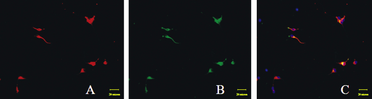
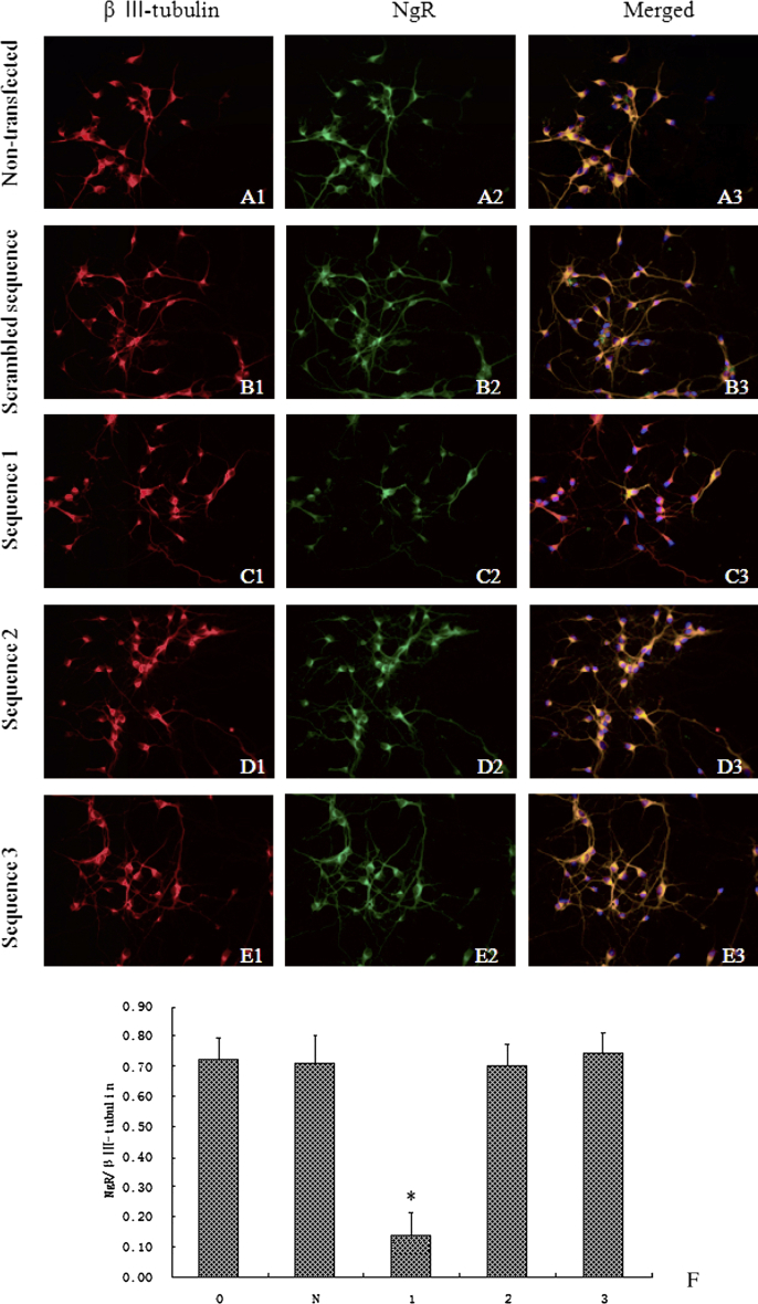
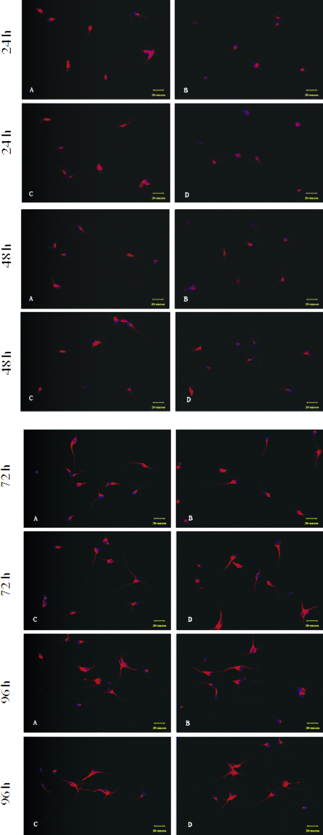
Methods: Three siRNA sequences against NgR were designed and transfected into cerebellar granule cells (CGCs) to screen for the most effcient sequence of NgR siRNA by using reverse transcription polymerase chain reaction (RT-PCR) and immunofluorescence staining. NgR siRNA sequence 1 was found the most efficient which was then transfected into the CGCs grown on CNS myelin substrate to observe its disinhibition for neurite outgrowth.

Results: Compared with the scrambled control sequence of siRNA, the NgR siRNA sequence 1 significantly decreased NgR mRNA level at 24 h and 48 h (p <0.05), which was recovered by 96 h after transfection. NgR immunoreactivity was also markedly reduced at 24 and 48 h after the transfection of siRNA sequence 1 compared with that before transfection (p<0.05). The NgR immunoreactivity was recovered after 72 h post-transfection. Moreover, the neurite outgrowth on the myelin substrate was greatly improved within 72 h after the transfection with siRNA sequence 1 compared with the scrambled sequence-transfected group or non-transfected group (p<0.05).

Conclusion: siRNA-mediated knockdown of NgR expression contributes to neurite outgrowth in vitro.


Figures

Fig. 1
Fig. 1
Light (A) and fluorescent (B) microscopic images of scrambled siRNA-transfected CGCs. Cells transfected with FAM-labeled siRNA are shown in green.
Fig. 2
Fig. 2
Representative fluorescence images of CGCs stained with the antibodies of β III-tubulin and NgR 24 h after plating. All β III-tubulin-positive cells (red) in panel A are also NgR-positive (green) in panel B. Panel C shows the merged images. Scale bar = 20 μm.
Fig. 3
Fig. 3
siRNA-mediated knockdown of NgR mRNA at 48 h after transfection. A: RT-PCR analysis of mRNA expression. B: Densitometric analysis demonstrates that the ratio of NgR density to that of HPRT is significantly decreased by siRNA sequence 1 transfection (*p < 0.05 vs non-transfected cells, n = 6 for each group). There are no significant changes in NgR mRNA levels in the cells transfected with siRNA sequence 2 or 3, or the scrambled sequence (p > 0.05 vs non-transfected cells). 0: non-transfected cells; N: scrambled control sequence; 1: siRNA sequence 1; 2: siRNA sequence 2; 3: siRNA sequence.
Fig. 4
Fig. 4
Time course of siRNA-mediated knockdown of NgR mRNA. A: RT-PCR analysis of mRNA expression. B: Quantification of NgR/HPRT by image analysis. The data show that the maximal inhibition of NgR mRNA is achieved at 24 h post-transfection (*p < 0.05 vs control). Inhibition is maintained for approximately 72 h, then the level of NgR mRNA is restored to that of the non-transfected cells.
Fig. 5
Fig. 5
A–E: NgR expression is suppressed by siRNA sequence 1 at 48 h after transfection. The cells, non-transfected or transfected with siRNA of sequence 1, 2, or 3, or scrambled sequence, are stained with β III-tubulin and NgR antibodies. F: Densitometric analysis shows that transfection of neurons with siRNA sequence 1 results in significant suppression in NgR (*p < 0.05 vs non-transfected cells), but no significant changes are detected in cells transfected with sequence 2 or 3, or the scrambled sequence (p > 0.05 vs non-transfected cells). 0: non-transfected cells; N: scrambled sequence; 1: siRNA sequence 1; 2: siRNA sequence 2; 3: siRNA sequence 3.
Fig. 6
Fig. 6
Microscopic images of double immunofluorescence stained neurons for β III-tubulin (red) and NgR (green) at different time points after transfection.
Fig. 7
Fig. 7
Effects of siRNA transfection on neurite outgrowth of CGCs in different culture substrates. Following plating, neurite outgrowth of the three groups treated with myelin membrane proteins is inhibited to some extent; however, significant promotion of neurite outgrowth is observed in group C (*p < 0.05 vs groups B and D). By 96 h, the difference among the four groups has become insignificant. A: noninhibitory culture; B: myelin membrane proteins; C: myelin membrane proteins + siRNA; D: myelin membrane proteins + scrambled sequence.
Fig. 8
Fig. 8
Microscopic images of immunofluorescence staining of neurons for β III-tubulin at 24–96 h after transfection. A: noninhibitory culture; B: myelin; C: myelin + siRNA; D: myelin + scrambled sequence. Scale bar = 20 μm.

Similar articles

References

    1. Mukhopadhyay G., Doherty P., Walsh F.S. A novel role for myelin-associated glycoprotein as an inhibitor of axonal regeneration. Neuron. 1994;13:757–767. - PubMed
    1. Domeniconi M., Cao Z., Spencer T. Myelin-associated glycoprotein interacts with the Nogo66 receptor to inhibit neurite outgrowth. Neuron. 2002;35:283–290. - PubMed
    1. Borisoff J.F., Chan C.C., Hiebert G.W. Suppression of Rho-kinase activity promotes axonal growth on inhibitory CNS substrates. Mol Cell Neurosci. 2003;22:405–416. - PubMed
    1. Harvey P.A., Lee D.H., Qian F. Blockade of Nogo receptor ligands promotes functional regeneration of sensory axons after dorsal root crush. J Neurosci. 2009;29:6285–6295. - PMC - PubMed
    1. Berry M., Hunter A.S., Duncan A. Axon-glial relations during regeneration of axons in the adult rat anterior medullary velum. J Neurocytol. 1998;27:915–937. - PubMed

Publication types