

Cervical vertebral actinomycosis mimicking malignancy in a paediatric patient
- PMID: 27033296
- PMCID: PMC4840670
- DOI: 10.1136/bcr-2016-214757
Cervical vertebral actinomycosis mimicking malignancy in a paediatric patient
Abstract
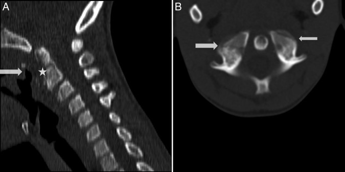
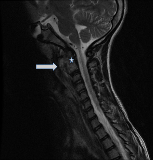
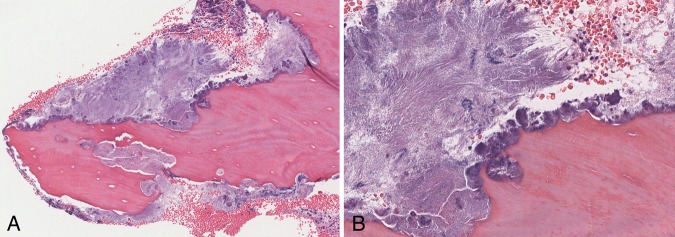
Actinomyces spp are found in the flora of the oral cavity and vagina and may cause infection with abscess formation and draining sinuses. Cervicofacial manifestations of actinomycosis involve head and neck soft tissue, however, spread to the cervical spine is rare. We report a case of an 8-year-old boy, presenting with neck pain for 1 month and denying a history of trauma or procedures. Radiography revealed an ulceration of the posterior oropharyngeal mucosa with a defect extending to the C1-C2 vertebra, mimicking a neoplastic process. The patient underwent laryngoscopy and multiple biopsies were taken from the ulcer and bone, showing severe osteomyelitis and intraosseous filamentous organisms, morphologically consistent with Actinomyces spp. The boy received long-term antibiotics with response to treatment. Actinomycosis has rarely been reported in the cervical vertebrae of paediatric patients. This should be considered as a differential diagnosis for such a presentation as prompt antibiotic treatment may be lifesaving.
2016 BMJ Publishing Group Ltd.
Figures

References
-
- Tortorici S, Burruano F, Buzzanca ML et al. . Cervico-facial actinomycosis: epidemiological and clinical comments. Am J Infect Dis 2008;4:204–8. 10.3844/ajidsp.2008.204.208 - DOI
-
- Bennett JE, Dolin R, Blaser MJ. Principles and practice of infectious diseases. Elsevier Health Sciences, 2014.
Publication types
MeSH terms
Substances
LinkOut - more resources
Full Text Sources
Other Literature Sources
Miscellaneous