

Phosphatidylethanolamine binding protein 4 (PEBP4) is a secreted protein and has multiple functions
- PMID: 27033522
- PMCID: PMC5336313
- DOI: 10.1016/j.bbamcr.2016.03.022
Phosphatidylethanolamine binding protein 4 (PEBP4) is a secreted protein and has multiple functions
Abstract
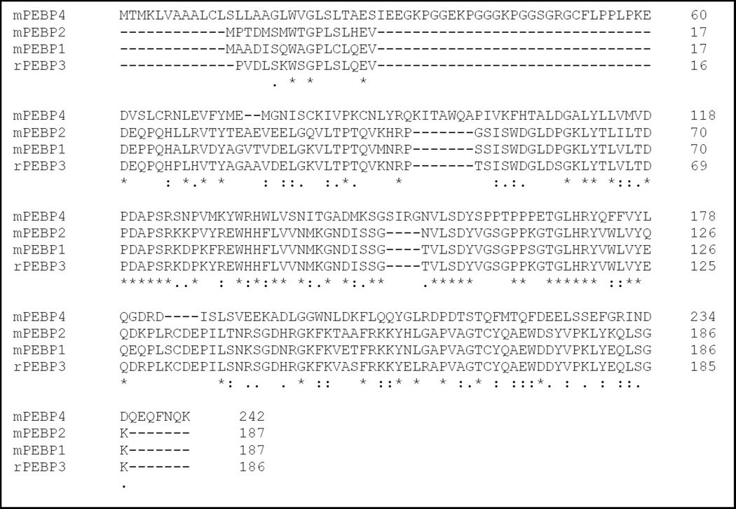
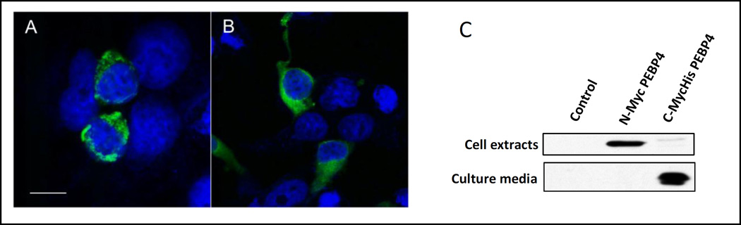
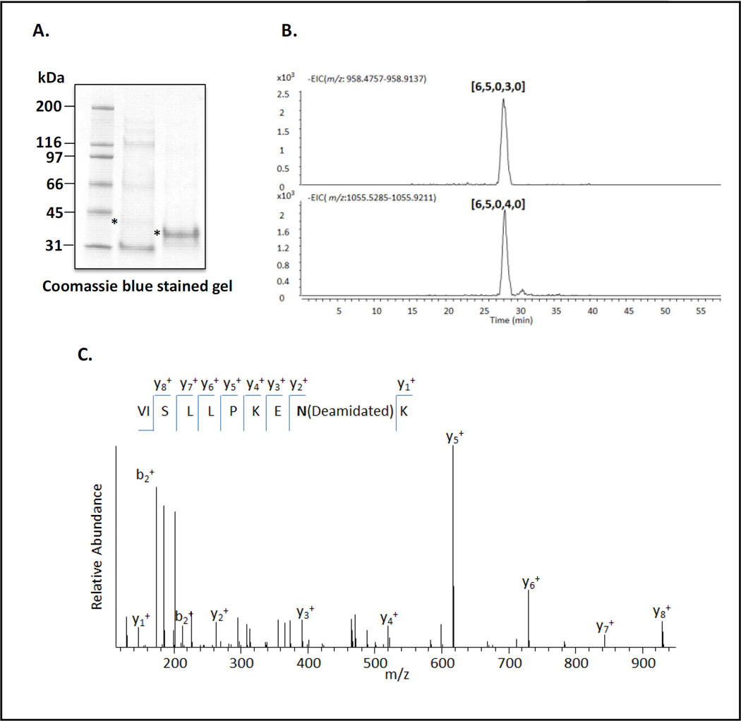
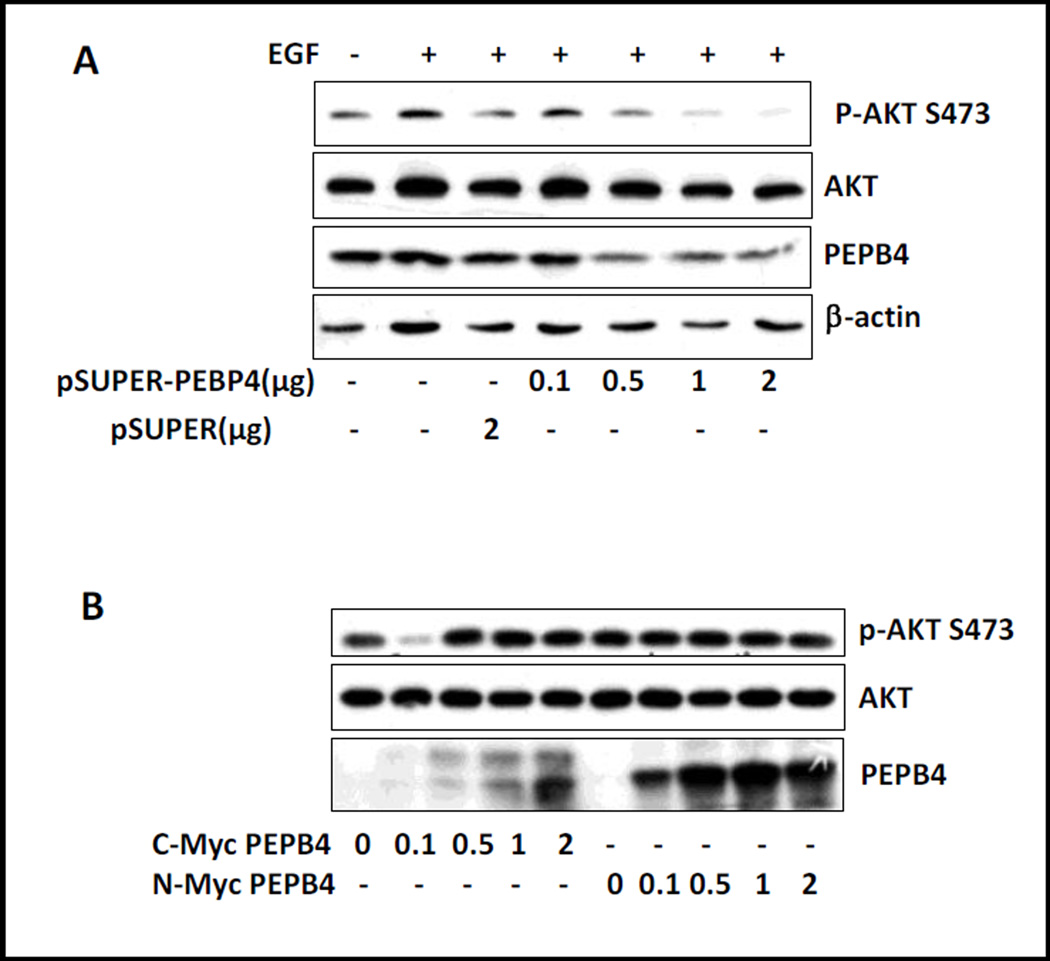
Phosphatidylethanolamine binding proteins (PEBP) represent a superfamily of proteins that are conserved from bacteria to humans. In mammals, four members have been identified, PEBP1-4. To determine the functional differences among PEBP1-4 and the underlying mechanism for their actions, we performed a sequence alignment and found that PEBP4 contains a signal peptide and potential glycosylation sites, whereas PEBP1-3 are intracellular proteins. To test if PEBP4 is secreted, we made constructs with Myc epitope at the amino (N) terminus or carboxyl (C) terminus to mask the signal sequence or keep it free, respectively. Our data revealed that both mouse and human PEBP4 were secreted when the epitope was tagged at their C-terminus. To our surprise, secretion was dependent upon the C-terminal conserved domain in addition to the N-terminal signal sequence. When the epitope was placed to the N-terminus, the recombinant protein failed to secrete and instead, was retained in the cytoplasm. Mass spectrometry detected asparagine (N)-glycosylation on the secreted PEBP4. Although overexpression of N-terminal tagged PEBP4 resulted in an inhibition of ERK activation by EGF, that with a C-terminal epitope tag did not have such an effect. Likewise, transfection of PEBP4 shRNA did not appear to affect ERK activation, suggesting that PEBP4 does not participate in the regulation of this pathway. In contrast, PEBP4 siRNA suppressed phosphorylation of Act at S473. Therefore, our results suggest that PEBP4 is a multifunctional protein and can be secreted. It will be important to investigate the mechanism by which PEBP4 is secreted and regulates cellular events.
Keywords: Act; ERK; Glycosylation; PEBP4; Secretion; Signal peptide.
Copyright © 2016 Elsevier B.V. All rights reserved.
Conflict of interest statement
The authors declare no conflicts of interest.
Figures


Similar articles
-
Regulation of human myoblast differentiation by PEBP4.EMBO Rep. 2009 Mar;10(3):278-84. doi: 10.1038/embor.2009.4. Epub 2009 Feb 6. EMBO Rep. 2009. PMID: 19197339 Free PMC article.
-
PEBP4 silencing inhibits hypoxia-induced epithelial-to-mesenchymal transition in prostate cancer cells.Biomed Pharmacother. 2016 Jul;81:1-6. doi: 10.1016/j.biopha.2016.03.030. Epub 2016 Apr 6. Biomed Pharmacother. 2016. PMID: 27261570
-
Role of the PEBP4 protein in the development and metastasis of gastric cancer.Oncotarget. 2017 Mar 14;8(11):18177-18184. doi: 10.18632/oncotarget.15255. Oncotarget. 2017. PMID: 28193908 Free PMC article.
-
The role of phosphatidylethanolamine-binding protein (PEBP) family in various diseases: Mechanisms and therapeutic potential.Prog Biophys Mol Biol. 2025 Jun;196:102-113. doi: 10.1016/j.pbiomolbio.2025.04.002. Epub 2025 Apr 10. Prog Biophys Mol Biol. 2025. PMID: 40220872 Review.
-
Understanding perspectives of signalling mechanisms regulating PEBP1 function.Cell Biochem Funct. 2016 Aug;34(6):394-403. doi: 10.1002/cbf.3198. Epub 2016 Jul 7. Cell Biochem Funct. 2016. PMID: 27385268 Review.
Cited by
-
Expression of uncharacterized male germ cell-specific genes and discovery of novel sperm-tail proteins in mice.PLoS One. 2017 Jul 25;12(7):e0182038. doi: 10.1371/journal.pone.0182038. eCollection 2017. PLoS One. 2017. PMID: 28742876 Free PMC article.
-
Characterization of PEBP-like Genes and Function of Capebp1 and Capebp5 in Fruiting Body Regeneration in Cyclocybe aegerita.J Fungi (Basel). 2024 Jul 31;10(8):537. doi: 10.3390/jof10080537. J Fungi (Basel). 2024. PMID: 39194863 Free PMC article.
-
The tobacco phosphatidylethanolamine-binding protein NtFT4 increases the lifespan of Drosophila melanogaster by interacting with the proteostasis network.Aging (Albany NY). 2022 Apr 8;14(7):2989-3029. doi: 10.18632/aging.204005. Epub 2022 Apr 8. Aging (Albany NY). 2022. PMID: 35396341 Free PMC article.
-
PEBP4 Directs the Malignant Behavior of Hepatocellular Carcinoma Cells via Regulating mTORC1 and mTORC2.Int J Mol Sci. 2022 Aug 8;23(15):8798. doi: 10.3390/ijms23158798. Int J Mol Sci. 2022. PMID: 35955931 Free PMC article.
-
Paraptosis: a unique cell death mode for targeting cancer.Front Pharmacol. 2023 Jun 15;14:1159409. doi: 10.3389/fphar.2023.1159409. eCollection 2023. Front Pharmacol. 2023. PMID: 37397502 Free PMC article. Review.
References
-
- Al-Mulla F, Bitar MS, Taqi Z, Yeung KC. RKIP: much more than Raf kinase inhibitory protein. J Cell Physiol. 2013;228(8):1688–1702. PubMed PMID: 23359513. - PubMed
-
- Frayne J, Ingram C, Love S, Hall L. Localisation of phosphatidylethanolamine-binding protein in the brain and other tissues of the rat. Cell Tissue Res. 1999;298(3):415–423. PubMed PMID: 10639732. - PubMed
-
- Katada E, Mitake S, Matsukawa N, Otsuka Y, Tsugu Y, Fujimori O, Ojika K. Distribution of hippocampal cholinergic neurostimulating peptide (HCNP)-like immunoreactivity in organs and tissues of young Wistar rats. Histochem Cell Biol. 1996;105(1):43–51. PubMed PMID: 8824905. - PubMed
-
- Hickox DM, Gibbs G, Morrison JR, Sebire K, Edgar K, Keah HH, Alter K, Loveland KL, Hearn MT, de Kretser DM, O'Bryan MK. Identification of a novel testis-specific member of the phosphatidylethanolamine binding protein family, pebp-2. Biol Reprod. 2002;67(3):917–927. PubMed PMID: 12193403. - PubMed
-
- Wang X, Li N, Liu B, Sun H, Chen T, Li H, Qiu J, Zhang L, Wan T, Cao X. A novel human phosphatidylethanolamine-binding protein resists tumor necrosis factor alpha-induced apoptosis by inhibiting mitogen-activated protein kinase pathway activation and phosphatidylethanolamine externalization. The Journal of biological chemistry. 2004;279(44):45855–45864. Epub 2004/08/11. PubMed PMID: 15302887. - PubMed
Publication types
MeSH terms
Substances
Grants and funding
LinkOut - more resources
Full Text Sources
Other Literature Sources
Molecular Biology Databases
Research Materials
Miscellaneous
