. 2016 Apr 29;473(2):565-71.
doi: 10.1016/j.bbrc.2016.03.122. Epub 2016 Mar 28.

Aldose reductase mediates retinal microglia activation

Affiliations

Aldose reductase mediates retinal microglia activation

Kun-Che Chang et al. Biochem Biophys Res Commun. .

Abstract

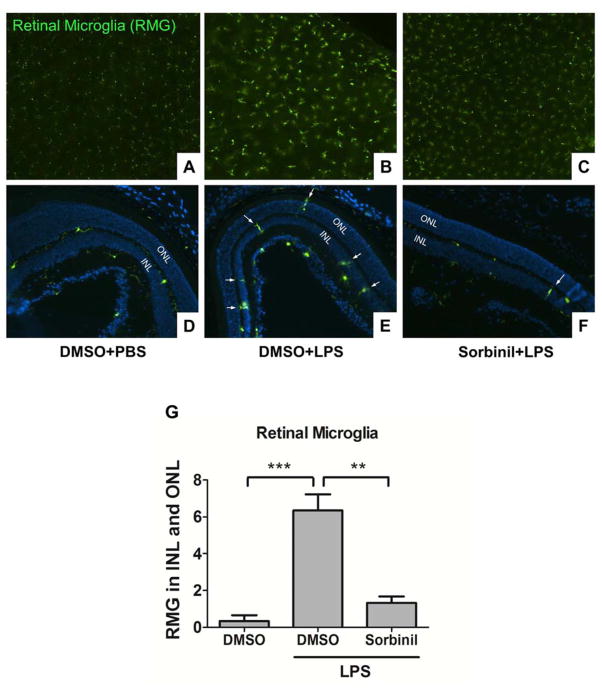
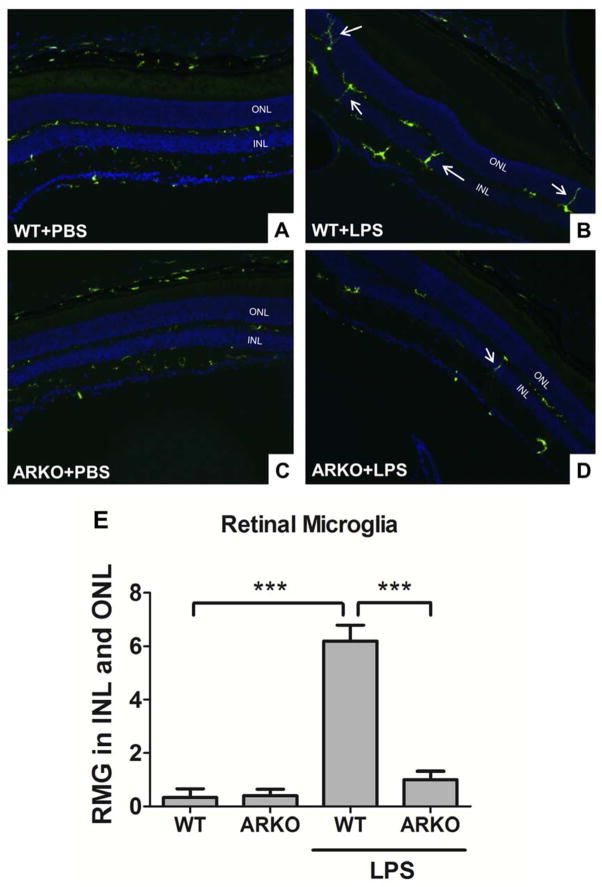
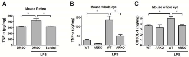
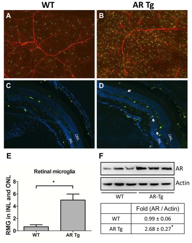
Retinal microglia (RMG) are one of the major immune cells in charge of surveillance of inflammatory responses in the eye. In the absence of an inflammatory stimulus, RMG reside predominately in the ganglion layer and inner or outer plexiform layers. However, under stress RMG become activated and migrate into the inner nuclear layer (INL) or outer nuclear layer (ONL). Activated RMG in cell culture secrete pro-inflammatory cytokines in a manner sensitive to downregulation by aldose reductase inhibitors. In this study, we utilized CX3CR1(GFP) mice carrying AR mutant alleles to evaluate the role of AR on RMG activation and migration in vivo. When tested on an AR(WT) background, IP injection of LPS induced RMG activation and migration into the INL and ONL. However, this phenomenon was largely prevented by AR inhibitors or in AR null mice, or was exacerbated in transgenic mice that over-express AR. LPS-induced increases in ocular levels of TNF-α and CX3CL-1 in WT mice were substantially lower in AR null mice or were reduced by AR inhibitor treatment. These studies demonstrate that AR expression in RMG may contribute to the proinflammatory phenotypes common to various eye diseases such as uveitis and diabetic retinopathy.

Keywords: Aldose reductase; Inflammation; Retinal microglia.


Conflict of interest statement

Conflict of Interest Statement

The authors declare that there are no conflicts of interest.

Figures

Fig. 1
Fig. 1. Aldose reductase inhibitor prevents microglia from LPS-induced activation in the retina
WT-CX3XR1GFP mice were injected with LPS (20 mg/kg body weight) in the presence of DMSO or Sorbinil (10 mg/kg body weight) for 24 h before sacrifice. Whole retinas from each group were mounted on slides (A–C) and cryo sections (D–F) were collected from each group stained with DAPI. GFP expression was used to indicate RMG. White arrows indicate migrated RMG in inner or outer nuclear layers. Statistic data was performed in a bar chart (G). Data shown are means ± SEM (N = 3). **P < 0.01, ***P < 0.005. INL, inner nuclear layer; ONL, outer nuclear layer.
Fig. 2
Fig. 2. Genetic ablation of aldose reductase prevents microglia from LPS-induced activation in the retina
WT-CX3XR1GFP mice (A and B) and ARKO-CX3XR1GFP mice (C and D) were injected with LPS (20 mg/kg body weight) for 24 h before sacrifice. Cryo sections were collected from each group and stained with DAPI. GFP expression was used to indicate RMG. White arrows indicate migrated RMG in inner or outer nuclear layers. Statistic data was performed in a bar chart (F). Data shown are means ± SEM (N = 3). ***P < 0.005. INL, inner nuclear layer; ONL, outer nuclear layer.
Fig. 3
Fig. 3. Pharmacological inhibition or genetic ablation of aldose reductase reduces LPS-induced cytokines expression in the retina and whole eye
WT mice (A) and ARKO mice (B and C) were injected with LPS (20 mg/kg body weight) for 24 h before sacrifice. Mouse retinas (A) or whole eyes (B and C) were lysed in RIPA buffer. TNF-α (A and B) and CX3CL-1 (C) were measured using ELISA kit. Data shown are means ± SEM (N = 3). *P < 0.05.
Fig. 4
Fig. 4. Overexpression of aldose reductase causes microglia activation in the retina
WT-CX3XR1GFP mice (A and C) and ARTg-CX3XR1GFP mice (B and D) were sacrificed at 3 months of age. Whole retinas from each group were mounted on slides with Isolectin IB4 staining (A and B) and cryo sections (C and D) were collected from each group stained with DAPI. GFP was spontaneously expressed indicating RMG. White arrows indicate migrated RMG in inner or outer nuclear layers. Statistic data was performed in a bar chart (E). Level of AR in retinas was measured by Western blot (F). Data shown are means ± SEM (N = 3). *P < 0.05. INL, inner nuclear layer; ONL, outer nuclear layer.

References

    1. del Rio-Hortega P. Art and artifice in the science of histology. 1933. Histopathology. 1993;22:515–525. - PubMed
    1. Vrabec F. Microglia in the monkey and rabbit retina. J Neuropathol Exp Neurol. 1970;29:217–224. - PubMed
    1. Ling EA. A light microscopic demonstration of amoeboid microglia and microglial cells in the retina of rats of various ages. Arch Histol Jpn. 1982;45:37–44. - PubMed
    1. Boya J, Calvo J, Prado A. The origin of microglial cells. J Anat. 1979;129:177–186. - PMC - PubMed
    1. Kettenmann H, Hanisch UK, Noda M, Verkhratsky A. Physiology of microglia. Physiol Rev. 2011;91:461–553. - PubMed

Publication types