. 2016 May;26(5):517-28.
doi: 10.1038/cr.2016.26. Epub 2016 Apr 1.

MLKL forms cation channels

Affiliations

MLKL forms cation channels

Bingqing Xia et al. Cell Res. 2016 May.

Abstract

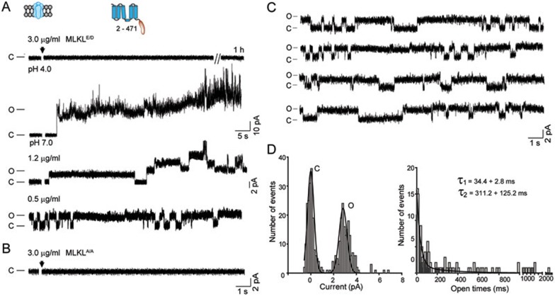
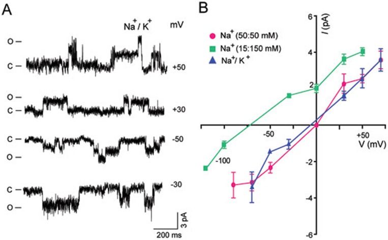
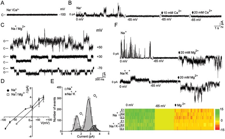
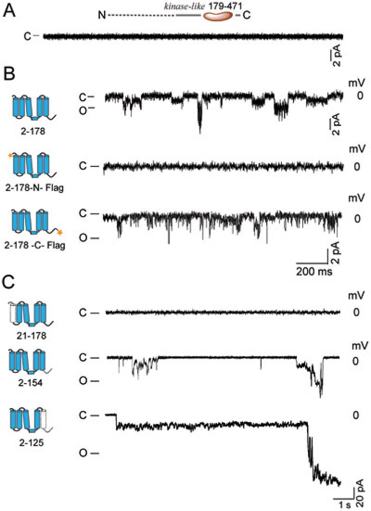
The mixed lineage kinase domain-like (MLKL) protein is a key factor in tumor necrosis factor-induced necroptosis. Recent studies on necroptosis execution revealed a commitment role of MLKL in membrane disruption. However, our knowledge of how MLKL functions on membrane remains very limited. Here we demonstrate that MLKL forms cation channels that are permeable preferentially to Mg(2+) rather than Ca(2+) in the presence of Na(+) and K(+). Moreover, the N-terminal domain containing six helices (H1-H6) is sufficient to form channels. Using the substituted cysteine accessibility method, we further determine that helix H1, H2, H3, H5 and H6 are transmembrane segments, while H4 is located in the cytoplasm. Finally, MLKL-induced membrane depolarization and cell death exhibit a positive correlation to its channel activity. The Mg(2+)-preferred permeability and five transmembrane segment topology distinguish MLKL from previously identified Mg(2+)-permeable channels and thus establish MLKL as a novel class of cation channels.


Figures

Figure 1
Figure 1
MLKL mediates typical single-channel currents in planar lipid bilayer. (A) MLKLE/D (3.0 μg/ml) did not induce current-like signals for 1 h in low pH solutions (pH = 4.0) (upper panel). In neutral solutions (pH = 7.0), 3.0 μg/ml MLKLE/D induced large currents while 1.2 μg/ml MLKLE/D induced multi-conductance step-like signals (middle panel) . A reduced amount of MLKLE/D(0.5 μg/ml) induced typical single-channel currents (lower panel). Protein was added to the cis side of the symmetric 50 mM NaCl solutions, and the membrane potential was clamped at +50 mV (trans side). The cis side indicates the side the protein was added and the membrane potential represents the voltage potential at the trans side. pH was buffered to 7 for all solutions used in this study except otherwise stated. Inset: the schematic representation of bilayer lipid recording (left) and the purified MLKL (right). (B) MLKLA/A(3.0 μg/ml) did not induce step-like current signals. (C) A 70-second continuous recording of single-channel currents. (D) Left panel: all-points amplitude histogram generated from the continuous single-channel recording of C. The two peaks represent the current levels of the closed (C, left) and open (O, right) channel. The line represents a Gaussian fit to the binned data (bin width = 0.125 pA). Right panel: open-time histograms generated from more than 900 events. Lines are exponential fits to the binned data (bin width = 0.75 ms) from each data set (n > 3).
Figure 2
Figure 2
Cation selectivity of MLKL channels. (A) Representative traces of MLKLE/D channels in Na+/K+ mixture solutions under indicated membrane potentials. (B) Current-voltage (I-V) plots demonstrating cation selectivity. The asymmetric (green squares) 15:150 mM (cis:trans) Na+ solutions did not affect the linear I-V relationship but left-shifted the reversal potential to −63.7 mV. With the addition of 150:15mM (cis:trans) K+ solutions to the Na+ gradient, the reversal potential shifted to −6.0 mV from −63.7 mV, indicating that the MLKLE/D channels cannot distinguish Na+ and K+. The single-channel conductance in the symmetric Na+(magenta circles), the asymmetric Na+(green squares) and the Na+/K+ mixture (blue triangles) solutions was 45.9 ± 2.7, 36.7 ± 3.6, and 47.0 ± 8.7 pS, respectively (n > 4 for each group).
Figure 3
Figure 3
MLKL forms Mg2+-preferred channels. (A) MLKLE/D did not induce currents in the presence of Na+/Ca2+ mixture solutions. The cis side solution contained 15 mM Na+ and 10 mM Ca2+. The trans side solution contained 150 mM Na+ and 0.1 mM Ca2+. (B) Representative step-like currents in the asymmetric 15:150 mM (cis:trans) Na+ solutions indicate the formation of MLKLE/D channels in the lipid membrane. The holding potential was next changed to −65 mV, the Na+ equilibrium potential, to eliminate the Na+ signals. Ca2+ was subsequently added to the cis side to a final concentration of first, 10 mM, and then, 20 mM. (C) Representative traces of MLKLE/Din the Na+/Mg2+ mixture solutions under the indicated holding potentials. The cis side solution contained 15 mM Na+ and 10 mM Mg2+. The trans side solution contained 150 mM Na+ and 0.1 mM Mg2+. (D) Current-voltage plots of MLKLE/D channels in the indicated solutions. Erev indicates the reversal potential of the pre-mixed Na+/Mg2+ solutions given that the permeability of Mg2+ and Na+ are equal. (E) All-points amplitude histogram of the Mg2+ currents generated from the recordings presented in B. 'O1' and 'O2' indicate two open states of MLKL channels in indicated solutions. (F) Mg2+ currents recorded in the Na+ or Na+/K+ solutions. The heat map shows the transition of the currents in three individual experiments in Na+ or Na+/K+ solutions. E1, E2 and E3 indicate three independent experiments.
Figure 4
Figure 4
MLKL N-terminal domain suffices to form channels. (A) C-terminal kinase-like domain (MLKL179-471) did not induce step-like currents. (B) N-terminal domain (MLKL2-178) sufficed to mediate step-like currents. (C) Representative traces induced by indicated truncations of the N-terminal domain. The truncated parts are indicated using dotted lines. All recordings were performed in asymmetric 15:150 mM (cis:trans) Na+ solutions (n > 4 for each group).
Figure 5
Figure 5
Identification of transmembrane segments. (A) Left: schematic representation of the residues examined and the corresponding six α-helices revealed by the solution MLKL structure. The dotted lines indicate the hypothetical lipid bilayer. MTSET is applied from either the cis or trans side. Mutations for MTSET test from cis (small lilac balls) or trans (small yellow balls) side are indicated. Right: topology of one subunit of MLKL channel. The dotted lines and gray circles represent the cellular membrane. The C-terminal kinase-like domain is colored in pink. (B) Responses of wild-type MLKL2-178 to MTSET. The black arrow indicates the application of MTSET. (C) Responses of indicated mutants to MTSET. The currents were recorded in asymmetric 15:150 mM (cis:trans) Na+ solutions (n > 4 for each group). MTSET (100 μM) was applied either in cis or trans side as described in Materials and Methods.
Figure 6
Figure 6
The correlation between channel activity and MLKL-induced cell death. (A) Representative whole-cell currents induced by the expression of indicated constructs. The inset shows the protocol applied to elicit the currents. The currents elicited by +60 mV are used to calculate the current density. Inset: schematic representation of the whole-cell recording. (B) Current density of the currents induced by the constructs shown in A (n > 8). (C) Current-voltage (I-V) plots. For clarity, only the I-V plots of MLKL/RIP3, MLKLE/D and MLKLA/A are shown (n > 8). (D) Time-dependent cell death (red line) and membrane potential depolarization (black line) induced by coexpressing MLKL/RIP3. Time (h) indicates the time after transfection. (E) The correlation between the current density, depolarization of the membrane potential and cell death induced by the indicated constructs. The parameters were measured 16-20 h after transfection. 'M/R' indicates MLKL/RIP3; 'E/D' indicates MLKLE/D; 'A/A' indicates MLKLA/A.

Comment in

References

    1. Pasparakis M, Vandenabeele P. Necroptosis and its role in inflammation. Nature 2015; 517:311–320. - PubMed
    1. Sun L, Wang X. A new kind of cell suicide: mechanisms and functions of programmed necrosis. Trends Biochem Sci 2014; 39:587–593. - PubMed
    1. Grooten J, Goossens V. Vanhaesebroeck, et al. Cell membrane permeabilization and cellular collapse, followed by loss of dehydrogenase activity: early events in tumour necrosis factor-induced cytotoxicity. Cytokine 1993; 5:546–555. - PubMed
    1. Laster SM, Wood JG, Gooding LR. Tumor necrosis factor can induce both apoptic and necrotic forms of cell lysis. J Immunol 1988; 141:2629–2634. - PubMed
    1. Vercammen D, Beyaert R, Denecker G, et al. Inhibition of caspases increases the sensitivity of L929 cells to necrosis mediated by tumor necrosis factor. J Exp Med 1998; 187:1477–1485. - PMC - PubMed

Publication types