. 2016 May 27;291(22):11551-65.
doi: 10.1074/jbc.M116.719138. Epub 2016 Mar 31.

Molecular Basis of the Ligand Binding Specificity of αvβ8 Integrin

Affiliations

Molecular Basis of the Ligand Binding Specificity of αvβ8 Integrin

Akio Ozawa et al. J Biol Chem. .

Abstract

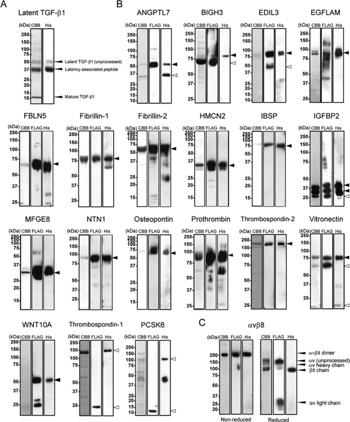
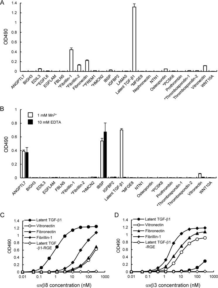
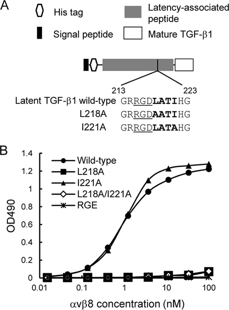
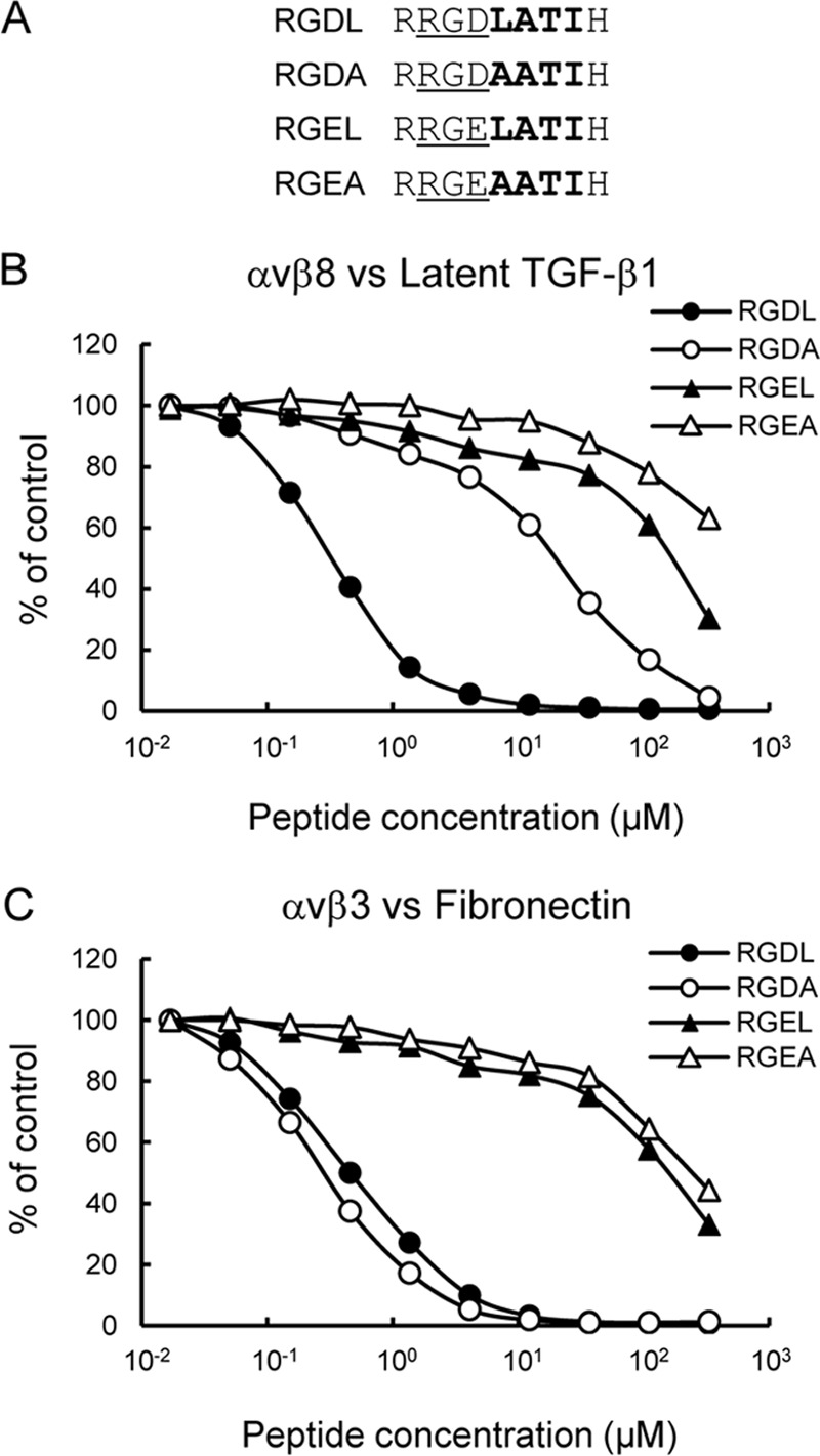
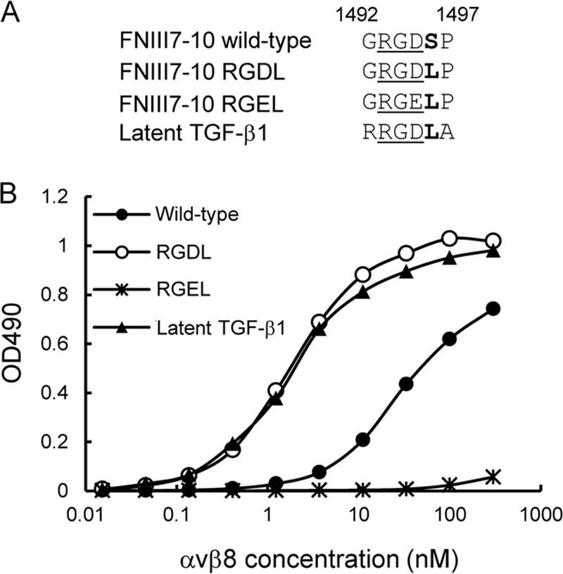
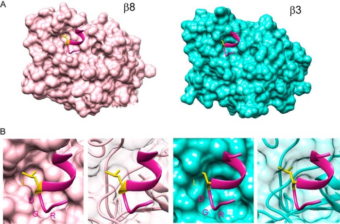
αvβ8 is an integrin that recognizes an Arg-Gly-Asp (RGD) motif and interacts with fibronectin, vitronectin, and latent TGF-β1. We comprehensively determined the binding activity of the αvβ8 integrin toward 25 secreted proteins having an RGD motif. The αvβ8 integrin strongly bound to latent TGF-β1 but showed marginal activity for other RGD-containing proteins, including fibronectin and vitronectin. Site-directed mutagenesis of latent TGF-β1 demonstrated that the high affinity binding of αvβ8 integrin to latent TGF-β1 was defined by Leu-218 immediately following the RGD motif within the latency-associated peptide of TGF-β1. Consistent with the critical role of Leu-218 in latent TGF-β1 recognition by αvβ8 integrin, a 9-mer synthetic peptide containing an RGDL sequence strongly inhibited interactions of latent TGF-β1 with αvβ8 integrin, whereas a 9-mer peptide with an RGDA sequence was ∼60-fold less inhibitory. Because αvβ3 integrin did not exhibit strong binding to latent TGF-β1 or distinguish between RGDL- and RGDA-containing peptides, we explored the mechanism by which the integrin β8 subunit defines the high affinity binding of latent TGF-β1 by αvβ8 integrin. Production of a series of swap mutants of integrin β8 and β3 subunits indicated that the high affinity binding of αvβ8 integrin with latent TGF-β1 was ensured by interactions between the Leu-218 residue and the β8 I-like domain, with the former serving as an auxiliary recognition residue defining the restricted ligand specificity of αvβ8 integrin toward latent TGF-β1. In support of this conclusion, high affinity binding toward the αvβ8 integrin was conferred on fibronectin by substitution of its RGDS motif with an RGDL sequence.

Keywords: RGD motif; cell adhesion; extracellular matrix; fibronectin; integrin; transforming growth factor beta (TGF-β).


Figures

FIGURE 1.
FIGURE 1.
Purification of recombinant RGD proteins and αvβ8 integrin. Purified latent TGF-β1 (A), other recombinant RGD proteins (B), and αvβ8 integrin (C) were subjected to SDS-PAGE on 5–20% gradient gels (ANGPTL7, EDIL3, PCSK6, and latent TGF-β1), 8% gels (EGFLAM, BIGH3, FBLN5, IGFBP2, NTN1, prothrombin, thrombospondin-2, vitronectin, WNT10A, thrombospondin-1, and αvβ8 integrin), or 12% gels (fibrillin-1, fibrillin-2, HMCN2, IBSP, MFGE8, and osteopontin) under reducing conditions, followed by Coomassie Brilliant Blue staining (left), immunoblotting with an anti-FLAG monoclonal antibody (middle), or with an anti-penta-His monoclonal antibody (right), except for αvβ8 integrin that was analyzed under both reducing and non-reducing conditions. Molecular masses are indicated on the left of panels. Arrowheads indicate predicted molecular size of full-length (close) or processed form (open) of each recombinant protein.
FIGURE 2.
FIGURE 2.
Binding activities of αvβ8 integrin toward 25 RGD proteins. A, microtiter plates were coated with RGD proteins (10 nm) and then incubated with αvβ8 integrin (10 nm) in the presence of 1 mm Mn2+. The bound integrins were quantified using a biotinylated anti-Velcro polyclonal antibody and HRP-conjugated streptavidin as described under “Experimental Procedures.” The amounts of integrin bound in the presence of 10 mm EDTA were used as negative controls and subtracted as background. The results represent S.E. of triplicate determinations. *, candidate proteins expressed as fragments containing an RGD motif. **, candidate proteins expressed as recombinant fragments fused to GST at their N termini. B, microtiter plates were coated with αvβ8(ΔHis) integrin (10 nm) and then incubated with RGD proteins (10 nm) in the presence of 1 mm Mn2+ or 10 mm EDTA. The bound RGD proteins were quantified using an HRP-conjugated anti-His6 antibody as described under “Experimental Procedures.” The results represent the means ± S.E. of triplicate determinations. C and D, titration curves of αvβ8 (left) and αvβ3 (right) integrins bound to latent TGF-β1 (closed circles), vitronectin (open circles), fibronectin (closed triangles), fibrillin-1 (closed diamonds), and the RGD → RGE substitution mutant of latent TGF-β1 (open diamonds). The results represent the means of three independent determinations. Bound integrins were quantified as described under “Experimental Procedures.”
FIGURE 3.
FIGURE 3.
Effect of alanine substitutions within the LATI sequence on αvβ8 integrin binding activity to latent TGF-β1. A, schematic of full-length TGF-β1 and the amino acid sequences of wild-type and alanine substitution mutants of latent TGF-β1. RGD motifs are underlined, and the following LATI sequences are shown in bold. B, titration curves of αvβ8 integrin bound to full-length TGF-β1 (wild-type, circles), L218A substitution mutant (L218A, squares), I221A substitution mutant (I221A, triangles), L218A/I221A double substitution mutant (L218A/I221A, open diamonds), and RGD → RGE mutant (RGE, asterisks). The assays were performed as described in the Fig. 2 legend. The results represent the means of three independent determinations.
FIGURE 4.
FIGURE 4.
Inhibition of αvβ8 integrin binding to latent TGF-β1 by synthetic peptides. A, amino acid sequences of the synthetic peptides tested. RGD motifs are underlined, and the following LATI sequences are shown in bold. B and C, integrins (10 nm) were incubated on microtiter plates coated with latent TGF-β1 (10 nm; B) or fibronectin (10 nm; C) in the presence of increasing concentrations of synthetic peptides. To prevent precipitation of the peptides, the integrin binding assays were performed in the presence of 10% DMSO. The amounts of bound integrins are shown as percentages relative to the control, in which integrins were incubated on latent TGF-β1- or fibronectin-coated plates in the presence of 10% DMSO. The results represent the means of three independent determinations. Closed circles, RGDL (9-mer containing both RGD motif and Leu residue); open circles, RGDA (9-mer with the Leu → Ala substitution); closed triangles, RGEL (9-mer with RGD → RGE substitution); open triangles, RGEA (9-mer with RGDL → RGEA double substitution).
FIGURE 5.
FIGURE 5.
Effect of leucine substitution for the serine residue immediately after the RGD motif in the 10th FNIII domain of fibronectin on its αvβ8 integrin binding activity. A, amino acid sequences of wild-type and leucine-substituted mutants of FNIII7–10. RGD motifs are underlined, and the subsequent residues are shown in bold. B, titration curves of αvβ8 integrin bound to wild-type FNIII7–10 (closed circles), RGDL mutant (open circles), RGEL mutant (asterisks), and latent TGF-β1 (triangles). The assays were performed as described in the legend for Fig. 2. The results represent the means of three independent determinations.
FIGURE 6.
FIGURE 6.
Ligand-binding specificities of domain swap mutants of αvβ8 and αvβ3 integrins. A, schematic of the ectodomain of integrin (left) and representations of the β8/β3 swap mutants (right). The β8- and β3-derived domains are represented by open boxes and closed boxes, respectively. The β8 hybrid domain is represented by dotted boxes. B, binding activities of domain swap mutants of αvβ8 and αvβ3 integrins toward latent TGF-β1, fibronectin, vitronectin, and fibrillin-1. C, titration curves of swap mutants bound to latent TGF-β1. Increasing concentrations of αvβ8 integrin (closed circles), αvβ3 integrin (closed squares), αvβ3–8BI/HYB (closed triangles), αvβ3–8BI (open squares), and αvβ8–3BI (open circles) were allowed to bind to microtiter plates coated with latent TGF-β1 in the presence of 1 mm MnCl2. Bound integrins were quantified as described under “Experimental Procedures.” The results represent the means of three independent determinations. Apparent dissociation constants of recombinant integrins are summarized in Table 3. D, inhibition of αvβ3–8BI binding to latent TGF-β1 by synthetic peptides. The assays were performed as described in the Fig. 4 legend. The results represent the means of three independent determinations. Closed circles, RGDL (9-mer containing both RGD motif and Leu residue); open circles, RGDA (9-mer with the Leu → Ala substitution); closed triangles, RGEL (9-mer with RGD → RGE substitution); open triangles, RGEA (9-mer with RGDL → RGEA double substitution).
FIGURE 7.
FIGURE 7.
Ligand-binding specificities of DLL swap mutants. A, schematic of the head region of integrin (left) and amino acid sequences of the DLL regions of the β8 and β3 subunits and their swap mutants (right). Swapped amino acids between the β8 and β3 subunits are indicated in the boxed area. B, binding activities of DLL swap mutants of αvβ8 and αvβ3 integrins toward latent TGF-β1, fibronectin, vitronectin, and fibrillin-1. C, titration curves of DLL swap mutants bound to latent TGF-β1 (left), fibronectin (middle), and vitronectin (right). Increasing concentrations of αvβ8 integrin (closed circles), αvβ3 integrin (closed squares), αvβ3–8DLL (open squares), and αvβ8–3DLL (open circles) were allowed to bind to microtiter plates coated with latent TGF-β1, fibronectin, or vitronectin in the presence of 1 mm MnCl2. Bound integrins were quantified as described under “Experimental Procedures.” The results represent the means of three independent determinations. Apparent dissociation constants of recombinant integrins are summarized in Table 3.
FIGURE 8.
FIGURE 8.
Inhibition of DLL swap mutant binding to latent TGF-β1 by synthetic peptides. αvβ8–3DLL (A) and αvβ3–8DLL (B) mutants (10 nm) were incubated on microtiter plates coated with latent TGF-β1 (10 nm) in the presence of increasing concentrations of synthetic peptides. To prevent precipitation of the peptides, the integrin binding assays were performed in the presence of 10% DMSO. The amounts of bound integrins are shown as percentages relative to the control, in which integrins were incubated on latent TGF-β1-coated plates in the presence of 10% DMSO. The results represent the means of three independent determinations. Closed circles, RGDL (9-mer containing both RGD motif and Leu residue); open circles, RGDA (9-mer with the Leu → Ala substitution); closed triangles, RGEL (9-mer with RGD → RGE substitution).
FIGURE 9.
FIGURE 9.
Predicted structures of the head region of αvβ8 integrin with an 11-mer peptide containing RGD motif and Leu residue. A, ribbon models of β I-like domain of αvβ8 integrin (left) and αvβ3 integrin (right) with 11-mer peptide HGRGDLGRLKK derived from latent TGF-β3 were created using the crystal structure of αvβ6 integrin with this peptide (Protein Data Bank code 4UM9) as the template. The models were predicted with the Swiss-Model and fine-tuned by energy minimization with UCSF Chimera. β I-like domains of β8 and β3 are shown in pink and cyan, respectively. 11-mer peptides are colored in violet red, and the Leu residue immediately following the RGD motif is shown with side chain in yellow. B, molecular surfaces of the β I-like domain of integrin β8 and β3 subunits with the 11-mer peptide containing RGD motif and Leu residue were generated with the Chimera. The β8 I-like domain is predicted to assume a structure of open conformation that allows the side chain of the Leu residue to fit into the β8 I-like domain, although the β3 I-like domain is predicted to assume a closed structure, failing to accommodate the side chain of the Leu residue.

Similar articles

Cited by

References

    1. Hynes R. O. (2002) Integrins: bidirectional, allosteric signaling machines. Cell 110, 673–687 - PubMed
    1. Takagi J. (2007) Structural basis for ligand recognition by integrins. Curr. Opin. Cell Biol. 19, 557–564 - PubMed
    1. Pytela R., Pierschbacher M. D., and Ruoslahti E. (1985) A 125/115-kDa cell surface receptor specific for vitronectin interacts with the arginine-glycine-aspartic acid adhesion sequence derived from fibronectin. Proc. Natl. Acad. Sci. U.S.A. 82, 5766–5770 - PMC - PubMed
    1. Brooks P. C., Clark R. A., and Cheresh D. A. (1994) Requirement of vascular integrin αvβ3 for angiogenesis. Science 264, 569–571 - PubMed
    1. Delannet M., Martin F., Bossy B., Cheresh D. A., Reichardt L. F., and Duband J. L. (1994) Specific roles of the αVβ1, αVβ3 and αVβ5 integrins in avian neural crest cell adhesion and migration on vitronectin. Development 120, 2687–2702 - PMC - PubMed

Publication types

Associated data