Review
. 2016 Sep 15:1647:65-78.
doi: 10.1016/j.brainres.2016.03.036. Epub 2016 Mar 28.

Mechanisms of FUS mutations in familial amyotrophic lateral sclerosis

Affiliations
Review

Mechanisms of FUS mutations in familial amyotrophic lateral sclerosis

Yulei Shang et al. Brain Res. .

Abstract

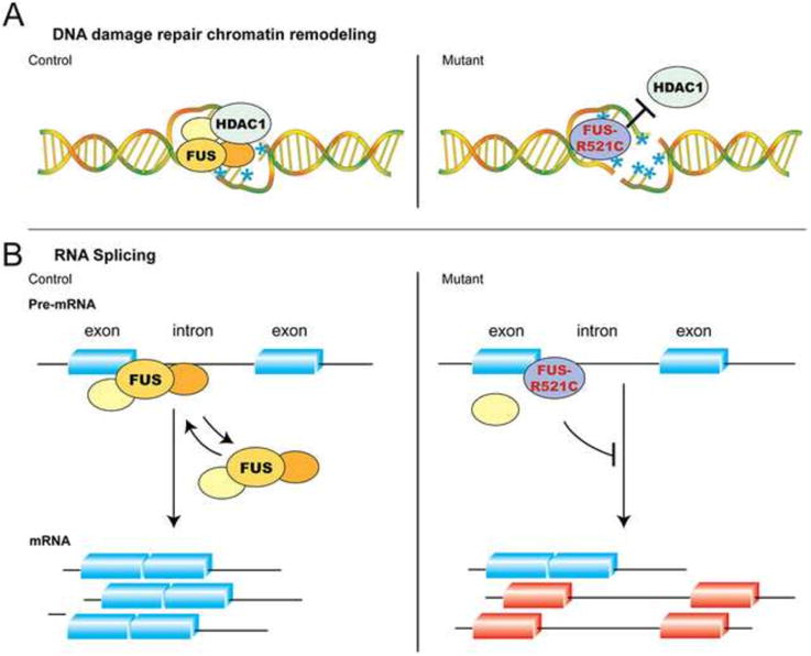
Recent advances in the genetics of amyotrophic lateral sclerosis (ALS) have provided key mechanistic insights to the pathogenesis of this devastating neurodegenerative disease. Among many etiologies for ALS, the identification of mutations and proteinopathies in two RNA binding proteins, TDP-43 (TARDBP or TAR DNA binding protein 43) and its closely related RNA/DNA binding protein FUS (fused in sarcoma), raises the intriguing possibility that perturbations to the RNA homeostasis and metabolism in neurons may contribute to the pathogenesis of these diseases. Although the similarities between TDP-43 and FUS suggest that mutations and proteinopathy involving these two proteins may converge on the same mechanisms leading to neurodegeneration, there is increasing evidence that FUS mutations target distinct mechanisms to cause early disease onset and aggressive progression of disease. This review focuses on the recent advances on the molecular, cellular and genetic approaches to uncover the mechanisms of wild type and mutant FUS proteins during development and in neurodegeneration. These findings provide important insights to understand how FUS mutations may perturb the maintenance of dendrites through fundamental processes in RNA splicing, RNA transport and DNA damage response/repair. These results contribute to the understanding of phenotypic manifestations in neurodegeneration related to FUS mutations, and to identify important directions for future investigations. This article is part of a Special Issue entitled SI:RNA Metabolism in Disease.

Keywords: Amyotrophic lateral sclerosis (ALS); DNA damage repair; Frontotemporal dementia (FTD); Fused in sarcoma (FUS); Low complexity domain; Prion-like property; RNA binding protein; RNA splicing.


Figures

Figure 1
Figure 1. The Age of Disease Onset in FALS Patients with FUS Mutations
(A) Meta-analyses of 154 FALS patients (either familial ALS inherited mutations or sporadic ALS with de novo mutations) show a predilection early disease onset. Compared to all FUS mutations and the most common mutations that occur in amino acid 521 (FUS-R521C), FUS-P525L mutation tends to occur in late teens and early 20's and represents a much more aggressive form of disease. (B) For sporadic ALS (SALS), about 35.9% and 34.9% of patients show disease onset in the range of 41-55 and 56-65 years old. In contrast, more than 60% of ALS patients with FUS mutations show disease onset before 40 years old. *Statistics for SALS have been adapted from the study by Testa and colleagues (Testa et al., 2004).
Figure 2
Figure 2. Schematic Diagrams of Genomic Organization of the Human FUS Gene, FUS Mutations Identified in ALS, and Functional Domains in FUS Proteins
The human FUS gene consists of 15 exons, spanning ∼14.9 Kb, and is located on chromosome 16p11.2. The FUS mRNA transcripts are predicted to contain a 3,433 bp 3′UTR, which has been recently shown to contain 4 disease-related variants. The full length human FUS protein contains 526 amino acids that can be further divided into several functional domains, including the “prion-like” or low complexity (LC) domain that contains the Q/G/S/Y-rich region (amino acids 1-165) and the G-rich region (amino acids 165-267), the Arginine-rich motif (RRM, amino acids 285-371), two Arg-Gly-Gly (RGG)-repeat regions (amino acids 371-422 and 453-501), interrupted by a Cys2-Cys2 zinc-finger motif (ZNF)(amino acids 422-453), and a non-conventional nuclear localization signal (NLS)(amino acids 510-526). The majority of FALS-related mutations are more commonly found in (1) the G-rich region, (2) the 2nd RGG region and (3) the NLS. Additional structural and functional domains in FUS include the prion-like domains and the HDAC1-interacting domains.
Figure 3
Figure 3. Dendritic and Synaptic Phenotypes Caused by FUS Mutations
Neurolucida tracing shows that the dendritic arbors in control motor neurons, highlighted by Golgi staining techniques, had 6 to 8 intersections per radial distance within 100 μm from the cell body, followed by a gradual reduction in the number of dendritic arbors from 100 to 250 μm. Compared to the control, the number of dendritic arbors in the FUS-R521C motor neurons shows no change within the first 50 μm from the cell body, but a significant reduction is noted from 50 to 250 μm, resulting in reduced cumulative dendritic area. To determine if the dendritic phenotype in FUS-R521C spinal motor neurons affected synaptic connectivity, we use ChAT (green) and FUS immunostaining to characterize the density of synapses surrounding motor neurons (Betley et al., 2009). Our results show that FUS proteins are present primarily in neuronal nuclei, but also show extensive colocalization with ChAT+ boutons and synaptophysin-immunoreactive presynaptic terminals. Remarkably, the density of ChAT+ boutons and SIPT showed significant reductions in the anterior horn of FUS-R521C spinal cord. To further characterize the synaptic defects, we use electron microscopy (EM) to ascertain the morphology and density of synapses within 100 μm radius of the cell body of spinal motor neurons, and show that the cell bodies of control motor neurons are surrounded by synaptic terminals arranged as rosette-like structures (Betley et al., 2009). In contrast, the size of post-synaptic density and the number of synapse per unit area are reduced in FUS-R521C motor neurons. Similar dendrite and synaptic defects are also noted in the apical and basal dendrites of the pyramidal neurons in layer IV-V of the sensorimotor cortex (Qiu et al., 2014), and neurons in the entorhinal cortex (Huang et al., 2012).
Figure 4
Figure 4. Mechanisms of Wild Type and Mutant FUS in DNA Damage Repair/Response and RNA Splicing
(A) Wild type FUS is rapidly recruited to DNA damage foci caused by double-stranded breaks, where it interacts with chromatin remodeling factor HDAC1. Although FUS-R521C can still be recruited to DNA damage foci, it fails to interact with HDAC1 and PARP1. Due to the defects in DNA repair/response machinery, neurons in FUS-R521C transgenic mice show increased DNA damage (indicated by blue asterisks and the presence of double-stranded breaks). (B) In addition to its role in DNA damage repair, several lines of evidence indicate that FUS can also regulate pre-mRNA splicing. Results from CLIP-RT-PCR and protein-RNA interactions in EMSA assays show that both wild type FUS and FUS-R521C can interact with selective oligoribonucleotides from Bdnf exon-intron boundaries. Whereas the equilibrium of wild type FUS-RNA interactions appears to be more dynamic, FUS-R521C tends to form more stable protein-RNA complexes that are more difficult to dissociate. (Figure adapted from Qiu et al., 2014, with permissions from the Journal of Clinical Investigation.)

References

    1. Alami NH, Smith RB, Carrasco MA, Williams LA, Winborn CS, Han SS, Kiskinis E, Winborn B, Freibaum BD, Kanagaraj A, et al. Axonal transport of TDP-43 mRNA granules is impaired by ALS-causing mutations. Neuron. 2014;81:536–543. - PMC - PubMed
    1. Alberti S, Halfmann R, King O, Kapila A, Lindquist S. A systematic survey identifies prions and illuminates sequence features of prionogenic proteins. Cell. 2009;137:146–158. - PMC - PubMed
    1. An JJ, Gharami K, Liao GY, Woo NH, Lau AG, Vanevski F, Torre ER, Jones KR, Feng Y, Lu B, et al. Distinct role of long 3′ UTR BDNF mRNA in spine morphology and synaptic plasticity in hippocampal neurons. Cell. 2008;134:175–187. - PMC - PubMed
    1. Anderson AJ, Su JH, Cotman CW. DNA damage and apoptosis in Alzheimer's disease: colocalization with c-Jun immunoreactivity, relationship to brain area, and effect of postmortem delay. J Neurosci. 1996;16:1710–1719. - PMC - PubMed
    1. Arai T, Hasegawa M, Akiyama H, Ikeda K, Nonaka T, Mori H, Mann D, Tsuchiya K, Yoshida M, Hashizume Y, et al. TDP-43 is a component of ubiquitin-positive tau-negative inclusions in frontotemporal lobar degeneration and amyotrophic lateral sclerosis. Biochem Biophys Res Commun. 2006;351:602–611. - PubMed

Publication types