Review
. 2016 Nov-Dec;61(6):745-758.
doi: 10.1016/j.survophthal.2016.03.007. Epub 2016 Mar 29.

Optic disk drusen in children

Affiliations
Review

Optic disk drusen in children

Melinda Y Chang et al. Surv Ophthalmol. 2016 Nov-Dec.

Abstract

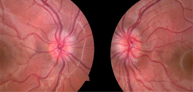
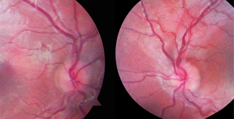
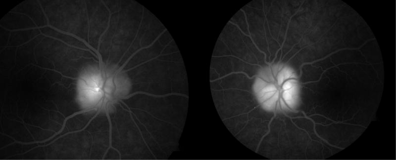
Optic disk drusen occur in 0.4% of children and consist of acellular intracellular and extracellular deposits that often become calcified over time. They are typically buried early in life and generally become superficial, and therefore visible, later in childhood, at the average age of 12 years. Their main clinical significance lies in the ability of optic disk drusen, particularly when buried, to simulate true optic disk edema. Misdiagnosing drusen as true disk edema may lead to an invasive and unnecessary workup for elevated intracranial pressure. Ancillary testing, including ultrasonography, fluorescein angiography, fundus autofluorescence, and optical coherence tomography, may aid in the correct diagnosis of optic disk drusen. Complications of optic disk drusen in children include visual field defects, hemorrhages, choroidal neovascular membrane, nonarteritic anterior ischemic optic neuropathy, and retinal vascular occlusions. Treatment options for these complications include ocular hypotensive agents for visual field defects and intravitreal anti-vascular endothelial growth factor agents for choroidal neovascular membranes. In most cases, however, children with optic disk drusen can be managed by observation with serial examinations and visual field testing once true optic disk edema has been excluded.

Keywords: children; choroidal neovascular membrane; optic disk drusen; pediatric; pseudopapilledema.


Figures

Figure 1
Figure 1
Comparison of optic disc in children with optic disc drusen and papilledema. A) Optic disc photos of a 10 year old boy with bilateral buried optic disc drusen. The disc margins are blurred, but there are no hemorrhages, exudates, or vessel obscuration. B) Optic disc photos of a 5 year old girl with mild papilledema due to increased intracranial pressure secondary to the use of exogenous growth hormone. Disc margins are blurred with mild obscuration of vessels, but no hemorrhages or exudates.
Figure 1
Figure 1
Comparison of optic disc in children with optic disc drusen and papilledema. A) Optic disc photos of a 10 year old boy with bilateral buried optic disc drusen. The disc margins are blurred, but there are no hemorrhages, exudates, or vessel obscuration. B) Optic disc photos of a 5 year old girl with mild papilledema due to increased intracranial pressure secondary to the use of exogenous growth hormone. Disc margins are blurred with mild obscuration of vessels, but no hemorrhages or exudates.
Figure 2
Figure 2
Characteristic visual field defects in a patient with bilateral optic disc drusen. The left eye has a small inferonasal scotoma, and the right eye has a predominantly nasal inferior arcuate defect.
Figure 3
Figure 3
Appearance of calcified optic disc drusen on ultrasonography. The calcified drusen produce a hyperechoic signal at the optic disc with posterior shadowing.
Figure 4
Figure 4
Fundus photography, autofluorescence, and fluorescein angiography in a 12 year old girl with optic disc drusen. A) Color fundus photography demonstrates blurred disc margins and superficial gliosis. B) Preinjection control photography shows hyperautofluorescence of optic disc drusen bilaterally. C) Late phase fluorescein angiography demonstrates nodular staining of the optic discs with no leakage.
Figure 4
Figure 4
Fundus photography, autofluorescence, and fluorescein angiography in a 12 year old girl with optic disc drusen. A) Color fundus photography demonstrates blurred disc margins and superficial gliosis. B) Preinjection control photography shows hyperautofluorescence of optic disc drusen bilaterally. C) Late phase fluorescein angiography demonstrates nodular staining of the optic discs with no leakage.
Figure 5
Figure 5
Optical coherence tomography of optic disc demonstrating drusen. The drusen appear as hyperreflective masses posterior to the outer plexiform and outer nuclear layers, with loss of the inner and outer segment photoreceptor junction.
Figure 6
Figure 6
Regressed juxtapapillary choroidal neovascular membrane secondary to optic disc drusen, with subretinal fibrosis and pigment mottling (Courtesy of Anthony C. Arnold, MD).

References

    1. Abegao Pinto L, Vandewalle E, Marques-Neves C, Stalmans I. Visual field loss in optic disc drusen patients correlates with central retinal artery blood velocity patterns. Acta Ophthalmol. 2014;92(4):e286–291. - PubMed
    1. Alkin Z, Ozkaya A, Yilmaz I, Yazici AT. A single injection of intravitreal ranibizumab in the treatment of choroidal neovascularisation secondary to optic nerve head drusen in a child. BMJ Case Rep. 2014 doi: 10.1136/bcr-2014-204456. - DOI - PMC - PubMed
    1. Anderson CJ, Zauel DW, Schlaeger TF, Meyer SM. Bilateral juxtapapillary subretinal neovascularization and pseudopapilledema in a three-year-old child. J Pediatr Ophthalmol Strabismus. 1978;15(5):296–299. - PubMed
    1. Antcliff RJ, Spalton DJ. Are optic disc drusen inherited? Ophthalmology. 1999;106(7):1278–1281. - PubMed
    1. Asensio-Sanchez VM, Trujillo-Guzman L. SD-OCT to distinguish papilledema from pseudopapilledema. Arch Soc Esp Oftalmol. 2015;90(10):481–483. - PubMed

Publication types

LinkOut - more resources