. 2016 May 3;7(18):26137-51.
doi: 10.18632/oncotarget.8433.

REST reduction is essential for hypoxia-induced neuroendocrine differentiation of prostate cancer cells by activating autophagy signaling

Affiliations

REST reduction is essential for hypoxia-induced neuroendocrine differentiation of prostate cancer cells by activating autophagy signaling

Tzu-Ping Lin et al. Oncotarget. .

Abstract

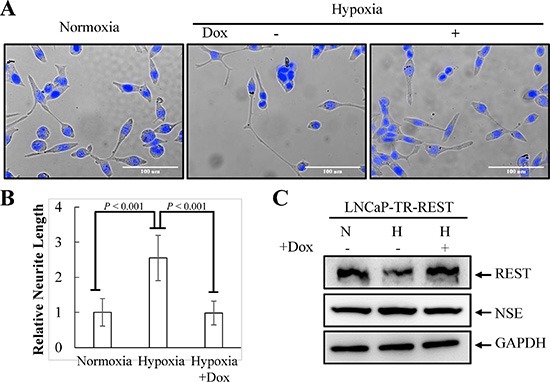
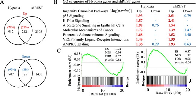
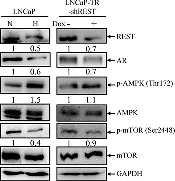
Prostate cancer (PCa) with neuroendocrine differentiation (NED) is tightly associated with hormone refractory PCa (HRPC), an aggressive form of cancer that is nearly impossible to treat. Determining the mechanism of the development of NED may yield novel therapeutic strategies for HRPC. Here, we first demonstrate that repressor element-1 silencing transcription factor (REST), a transcriptional repressor of neuronal genes that has been implicated in androgen-deprivation and IL-6 induced NED, is essential for hypoxia-induced NED of PCa cells. Bioinformatics analysis of transcriptome profiles of REST knockdown during hypoxia treatment demonstrated that REST is a master regulator of hypoxia-induced genes. Gene set enrichment analysis (GSEA) of hypoxia and REST knockdown co-upregulated genes revealed their correlation with HRPC. Consistently, gene ontology (GO) analysis showed that REST reduction potential associated with hypoxia-induced tumorigenesis, NE development, and AMPK pathway activation. Emerging reports have revealed that AMPK activation is a potential mechanism for hypoxia-induced autophagy. In line with this, we demonstrate that REST knockdown alone is capable of activating AMPK and autophagy activation is essential for hypoxia-induced NED of PCa cells. Here, making using of in vitro cell-based assay for NED, we reveal a new role for the transcriptional repressor REST in hypoxia-induced NED and characterized a sequential molecular mechanism downstream of REST resulting in AMPK phosphorylation and autophagy activation, which may be a common signaling pathway leading to NED of PCa.

Keywords: REST; autophagy; hypoxia; neuroendocrine differentiation; prostate cancer.


Conflict of interest statement

The authors disclose no potential conflicts of interest.

Figures

Figure 1
Figure 1. Hypoxia induces NED of LNCaP cells concomitant with down-regulation REST protein levels but not REST mRNA
(A) LNCaP cells were treated with hypoxia (2% O2) for 3 days. Representative photos of control and hypoxia-treated cells were stained with Hoechst. (B) The induced neurite length was assessed using brightfield microscopy images (40× magnification) and quantified by the average from 10 microscopic fields; bars, SD. (C) Total cell lysates (TCLs) were prepared from LNCaP cells treated as described in (A) for 1, 2 and 3 days and then immunoblotted to detect REST, AR, β-tubulin III, NSE, and β-TrCP. GAPDH was used as the loading control. (D) RT-qPCR analysis of total RNA from LNCaP cells treated as described in (A). The relative mRNA level of REST was normalized with B2M. Values from 3 independent experiments are reported as mean ± SD. (E) LNCaP cells were treated with hypoxia for 3 days in the presence or absence of 0.09 μM MG-132. The expression of REST was detected by immunoblotting using anti-REST antibody. GAPDH was used as the loading control.
Figure 2
Figure 2. Inhibition of neurite elongation by REST overexpression
(A) LNCaP-TR-REST cells were treated with 0.001 μg/ml Dox under normoxia or hypoxia (2% O2) conditions for 4 days. LNCaP-TR-REST cells without Dox treatment under normoxia was used as control. Representative photos of control and hypoxia-treated cells with or without REST overexpression were stained with Hoechst. (B) The neurite length was assessed using brightfield microscopy images (40× magnification) and quantified by the average from 10 microscopic fields; bars, SD. (C) The expression of REST and NSE under hypoxia treatment as described in (A) was confirmed using anti-REST and anti-NSE specific antibodies. GAPDH was used as the loading control.
Figure 3
Figure 3. Comparison of RNA-seq data sets between REST knockdown and hypoxia-treated LNCaP cells
(A) Venn diagrams depict the overlapped up- and down-regulated genes in REST knockdown and hypoxia-treated (2% O2) LNCaP cells. Total numbers of genes are listed inside the circles and the percentages of hypoxia-regulated genes are given. (B) Ingenuity canonical pathway (IPA) analysis of REST knockdown and hypoxia up- and down-regulated genes. –log (p value) > 1.3 was considered statistically significant and labeled in red. (C) The GSEA result showing the correlation of REST knockdown and hypoxia co-upregulated genes with PCa relapse (right panel) but not the malignance (left panel).
Figure 4
Figure 4. AMPK/mTOR pathway is activated by hypoxia treatment and REST knockdown
(A) LNCaP cells were treated with normoxia (N) or hypoxia (H) (2% O2) for 3 days (left panel). LNCaP-TR-shREST cells were treated with 1 μg/ml Dox for 3 days to knockdown REST (right panel). Total cell lysates (TLCs) were analyzed by immunoblotting using anti-REST, anti-AR, anti-p-AMPK, and anti-p-mTOR antibodies. Total AMPK and mTOR are used as controls. GAPDH was used as loading control. (B) The expression of each protein in three independent experiments was quantified; bars, SD.
Figure 5
Figure 5. AMPK pathway is essential for hypoxia-induced NED and autophagy activation
(A) LNCaP cells were treated with hypoxia (12% O2) for 3 days in the presence or absence of 2.5 μM compound C. Representative images of normoxia and hypoxia-treated cells with or without compound C were stained with Hoechst (left panel). The neurite length was assessed using brightfield microscopy images (40× magnification) and quantified by the average from 10 microscopic fields; bars, SD (right panel). (B) LNCaP-eGFP-LC3 cells were treated with hypoxia for 3 days in the presence or absence of 2.5 μM compound C. Following fixation, the cells were stained with Hoechst (Blue) and analyzed by fluorescence microscopy (FITC, 63× magnification) (left panel). The percentage of cells with punctate eGFP-LC3 expression was calculated using 10 microscopic fields and analyzed by MetaMorph software (right panel).
Figure 6
Figure 6. Hypoxia-induced autophagy activation of LNCaP cells
(A) LNCaP-eGFP-LC3 cells were treated with hypoxia (2% O2) for 3 days. Normoxia was used as control. Following fixation, the cells were stained with Hoechst (Blue) and analyzed by fluorescence microscopy (FITC, 63× magnification). (B) The percentage of cells with punctate eGFP-LC3 expression was calculated using 10 microscopic fields and analyzed by MetaMorph software. (C) TCLs were prepared from LNCaP cells treated with hypoxia for 1, 2 and 3 days and then immunoblotted to detect LC3. GAPDH was used as the loading control.
Figure 7
Figure 7. Chemical inhibition of autophagy flux by chloroquine (CQ) suppresses hypoxia-induced NED in LNCaP cells
(A) LNCaP cells were treated with hypoxia (2% O2) in the absence or presence of 50 μM CQ for 3 days. Following nuclear counterstaining with Hoechst (Blue), neurite structures were assessed by microscopy images (40× magnification). (B) Neurite lengths were quantified using 10 fields; bars, SD. (C) TLCs prepared from LNCaP cells in normoxia or treated with hypoxia in the absence or presence of 50 μM CQ for 3 days were analyzed by immunoblotting using the indicated antibodies.
Figure 8
Figure 8. Knockdown of beclin 1 suppresses hypoxia-induced NED
(A) LNCaP-TR-shBeclin1 cells were treated with 1 μg/ml Dox to induce beclin 1 knockdown for 4 days under normoxia and hypoxia (2% O2). Following nuclear counterstaining with Hoechst (Blue), the neurite structures were assessed by microscopy images (40×magnification). (B) The neurite length was quantified by 10 microscopic fields; bars, SD. (C) TLCs were prepared from LNCaP-TR-shBeclin1 cells treated as described in (A) and analyzed by immunoblotting using the indicated antibodies.
Figure 9
Figure 9. Knockdown of Atg5 suppresses hypoxia-induced NED
(A) LNCaP-TR-shAtg5 cells were treated with 1 μg/ml Dox to induce Atg5 knockdown for 4 days under normoxia and hypoxia (2% O2). Following nuclear counterstaining with Hoechst (Blue), the neurite structures were assessed by microscopy images (40× magnification). (B) Neurite lengths were quantified by 10 microscopic fields; bars, SD. (C) TLCs were prepared from LNCaP-TR-shAtg5 cells treated as described in (A) and analyzed by immunoblotting using the indicated antibodies.

References

    1. Wen S, Niu Y, Lee SO, Chang C. Androgen receptor (AR) positive vs negative roles in prostate cancer cell deaths including apoptosis, anoikis, entosis, necrosis and autophagic cell death. Cancer Treat Rev. 2014;40:31–40. - PMC - PubMed
    1. Aloysius H, Hu L. Targeted prodrug approaches for hormone refractory prostate cancer. Med Res Rev. 2015;35:554–585. - PubMed
    1. Conteduca V, Aieta M, Amadori D, De Giorgi U. Neuroendocrine differentiation in prostate cancer: current and emerging therapy strategies. Crit Rev Oncol Hematol. 2014;92:11–24. - PubMed
    1. Jin RJ, Wang Y, Masumori N, Ishii K, Tsukamoto T, Shappell SB, Hayward SW, Kasper S, Matusik RJ. NE-10 neuroendocrine cancer promotes the LNCaP xenograft growth in castrated mice. Cancer Res. 2004;64:5489–5495. - PubMed
    1. Burchardt T, Burchardt M, Chen MW, Cao Y, de la Taille A, Shabsigh A, Hayek O, Dorai T, Buttyan R. Transdifferentiation of prostate cancer cells to a neuroendocrine cell phenotype in vitro and in vivo. J Urol. 1999;162:1800–1805. - PubMed

MeSH terms

Substances

LinkOut - more resources