

Characterization of New Zealand White Rabbit Gut-Associated Lymphoid Tissues and Use as Viral Oncology Animal Model
- PMID: 27034393
- PMCID: PMC4816124
- DOI: 10.1093/ilar/ilw004
Characterization of New Zealand White Rabbit Gut-Associated Lymphoid Tissues and Use as Viral Oncology Animal Model
Abstract
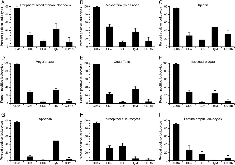
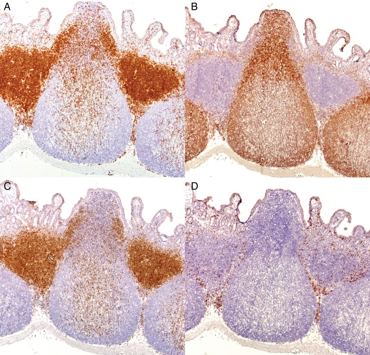
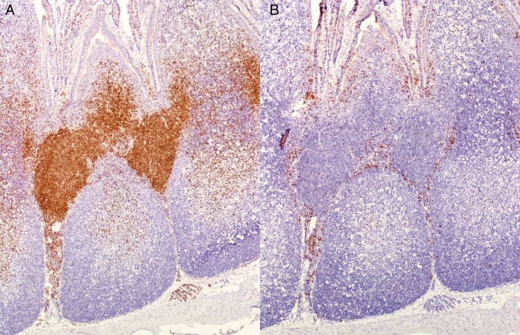
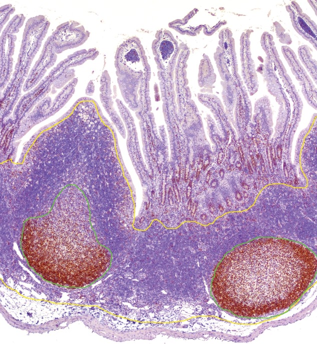
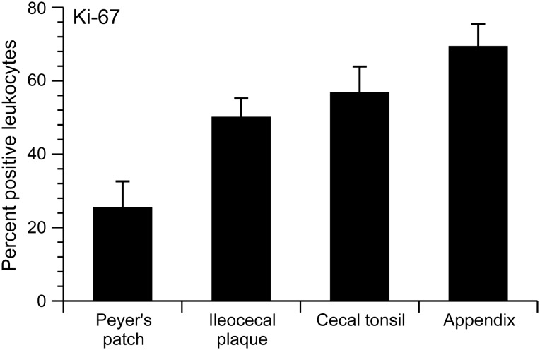
Rabbits have served as a valuable animal model for the pathogenesis of various human diseases, including those related to agents that gain entry through the gastrointestinal tract such as human T cell leukemia virus type 1. However, limited information is available regarding the spatial distribution and phenotypic characterization of major rabbit leukocyte populations in mucosa-associated lymphoid tissues. Herein, we describe the spatial distribution and phenotypic characterization of leukocytes from gut-associated lymphoid tissues (GALT) from 12-week-old New Zealand White rabbits. Our data indicate that rabbits have similar distribution of leukocyte subsets as humans, both in the GALT inductive and effector sites and in mesenteric lymph nodes, spleen, and peripheral blood. GALT inductive sites, including appendix, cecal tonsil, Peyer's patches, and ileocecal plaque, had variable B cell/T cell ratios (ranging from 4.0 to 0.8) with a predominance of CD4 T cells within the T cell population in all four tissues. Intraepithelial and lamina propria compartments contained mostly T cells, with CD4 T cells predominating in the lamina propria compartment and CD8 T cells predominating in the intraepithelial compartment. Mesenteric lymph node, peripheral blood, and splenic samples contained approximately equal percentages of B cells and T cells, with a high proportion of CD4 T cells compared with CD8 T cells. Collectively, our data indicate that New Zealand White rabbits are comparable with humans throughout their GALT and support future studies that use the rabbit model to study human gut-associated disease or infectious agents that gain entry by the oral route.
Keywords: gut-associated lymphoid tissue (GALT); human T cell leukemia/lymphoma virus 1 (HTLV-1); intraepithelial lymphocytes; lamina propria lymphocytes; lymphoid tissue; rabbit; review; small intestine.
© The Author 2016. Published by Oxford University Press on behalf of the Institute for Laboratory Animal Research. All rights reserved. For permissions, please email: journals.permissions@oup.com.
Figures
References
-
- Agace WW, Roberts AI, Wu L, Greineder C, Ebert EC, Parker CM. 2000. Human intestinal lamina propria and intraepithelial lymphocytes express receptors specific for chemokines induced by inflammation. Eur J Immunol 30:819–826. - PubMed
-
- Akagi T, Takeda I, Oka T, Ohtsuki Y, Yano S, Miyoshi I. 1985. Experimental infection of rabbits with human T-cell leukemia virus type 1. Jpn J Cancer Res 76:86–94. - PubMed
-
- Brandtzaeg P, Kiyono H, Pabst R, Russell MW. 2008. Terminology: Nomenclature of mucosa-associated lymphoid tissue. Mucosal Immunol 1:31–37. - PubMed
Publication types
MeSH terms
Grants and funding
LinkOut - more resources
Full Text Sources
Other Literature Sources
Research Materials
