. 2016 May;5(5):572-9.
doi: 10.5966/sctm.2015-0276. Epub 2016 Mar 31.

Modeling Cystic Fibrosis Using Pluripotent Stem Cell-Derived Human Pancreatic Ductal Epithelial Cells

Affiliations

Modeling Cystic Fibrosis Using Pluripotent Stem Cell-Derived Human Pancreatic Ductal Epithelial Cells

Senem Simsek et al. Stem Cells Transl Med. 2016 May.

Abstract

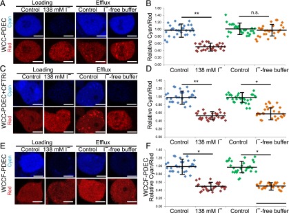
We established an efficient strategy to direct human pluripotent stem cells, including human embryonic stem cells (hESCs) and an induced pluripotent stem cell (iPSC) line derived from patients with cystic fibrosis, to differentiate into pancreatic ductal epithelial cells (PDECs). After purification, more than 98% of hESC-derived PDECs expressed functional cystic fibrosis transmembrane conductance regulator (CFTR) protein. In addition, iPSC lines were derived from a patient with CF carrying compound frameshift and mRNA splicing mutations and were differentiated to PDECs. PDECs derived from Weill Cornell cystic fibrosis (WCCF)-iPSCs showed defective expression of mature CFTR protein and impaired chloride ion channel activity, recapitulating functional defects of patients with CF at the cellular level. These studies provide a new methodology to derive pure PDECs expressing CFTR and establish a "disease in a dish" platform to identify drug candidates to rescue the pancreatic defects of patients with CF.

Significance: An efficient strategy was established to direct human pluripotent stem cells, including human embryonic stem cells (hESCs) and an induced pluripotent stem cell line derived from patients with cystic fibrosis (CF-iPSCs), to differentiate into pancreatic ductal epithelial cells (PDECs). After purification, more than 98% of hESC-PDECs derived from CF-iPSCs showed defective expression of mature cystic fibrosis transmembrane conductance regulator (CFTR) protein and impaired chloride ion channel activity, recapitulating functional pancreatic defects of patients with CF at the cellular level. These studies provide a new methodology for deriving pure PDECs expressing CFTR, and they establish a "disease-in-a-dish" platform for identifying drug candidates to rescue the pancreatic defects of these patients.

Keywords: Chloride ion efflux assay; Directed differentiation; Pancreatic defects of cystic fibrosis; Reprogramming.


Figures

Figure 1.
Figure 1.
Directed differentiation from hESCs to PDECs. (A): Scheme of the directed differentiation protocol. (B): Quantitative real-time polymerase chain reaction (qRT-PCR) analysis of HUES8-derived PDECs that were treated with 300 nM LDN193189, 10 ng/ml BMP2, 10 ng/ml BMP4, or 10 ng/ml BMP2 plus 10 ng/ml BMP4 during differentiation (n = 3 independent experiments). (C): Intracellular flow cytometry analysis of HUES8-derived PDECs that were treated with 10 ng/ml BMP4 from D16 to D27. (D): qRT-PCR analysis of HUES8-derived PDECs that were treated with or without 20 μM VPA from D16-D27 (n = 3 independent experiments). (E): Intracellular flow cytometry analysis of HUES8-derived PDECs that were treated with 10 ng/ml BMP4 plus 20 μM VPA from D16-D27 (n = 3 independent experiments). (F): qRT-PCR analysis of PDEC marker genes, including CFTR, CK19, CAII, and BEST1, in HUES8 cells, DE, PP, and PDECs (n = 3 independent experiments). (G): Immunocytochemistry analysis of HUES8-derived PDECs using CK19, CFTR, and SOX9 antibodies. Scale bars = 10 μM. Data are presented as mean ± SD; p values were calculated by unpaired two-tailed Student t test. ∗, p < .05; ∗∗, p < .01. Abbreviations: BMP, bone morphogenetic protein; CFTR, cystic fibrosis transmembrane conductance regulator; D, day; DE, definitive endoderm; DMEM, Dulbecco’s modified Eagle’s medium; DMEM+B27, Dulbecco’s modified Eagle’s medium supplemented with 1× B27; FBS, fetal bovine serum; hESC, human embryonic stem cell; n.s., no significant difference; PDEC, pancreatic ductal epithelial cell; PP, pancreatic progenitor; RPMI, Roswell Park Memorial Institute medium; SCC, side scatter; VPA, valproic acid.
Figure 2.
Figure 2.
Enrichment of CFTR-positive PDECs using a lentiviral vector containing CFTR-Puro-CMV-ClopHensor vector. Western blot (A), intracellular flow cytometry analysis (B), and quantitative real-time polymerase chain reaction analysis (C) of HUES8-derived PDECs before and after puromycin selection. n = 3 independent experiments in (C). Data are presented as mean ± SD; p values were calculated by unpaired two-tailed Student t test. ∗∗, p < .01. Abbreviations: CFTR, cystic fibrosis transmembrane conductance regulator; GAPDH, glyceraldehyde 3-phosphate dehydrogenase; PDEC, pancreatic ductal epithelial cell; SCC, side scatter; PDEC, pancreatic ductal epithelial cell; WCC, Weill Cornell control patient.
Figure 3.
Figure 3.
iPSC derivation and characterization. (A): WCCF-iPSC and WCC-iPSCs show typical embryonic stem cell-like colony morphology and express pluripotency markers. Scale bars = 50 μm. (B): The mutations of WCCF-iPSCs were confirmed using genomic DNA sequencing; WCC- and WCCF-iPSCs are capable of differentiating into PDX1-positive PPs (C) and CK19+/SOX9+ PDECs (n = 3 independent experiments) (D). Scale bars = 10 μm. (E): Quantification of CK19+/SOX9+ cells in WCC- and WCCF-iPSC-derived PDECs (n = 3 independent experiments). (F): Quantitative real-time polymerase chain reaction analysis of PDEC marker genes, including CFTR, CK19, CAII, and BEST1, in WCC-iPSC- and WCCF-iPSC-derived PDECs. Data are presented as mean ± SD; p values were calculated by unpaired two-tailed Student t test. Abbreviations: CFTR, cystic fibrosis transmembrane conductance regulator; DAPI, 4′,6-diamidino-2-phenylindole; iPSC, induced pluripotent stem cell; n.s., no significant difference; PDEC, pancreatic ductal epithelial cell; PP, pancreatic progenitor; WCC, Weill Cornell control patient; WCCF, Weill Cornell cystic fibrosis patient.
Figure 4.
Figure 4.
WCCF-induced pluripotent stem cell (iPSC)-derived pancreatic ductal epithelial cells (PDECs) show defective expression of mature CFTR protein. Immunocytochemistry (A) and Western blot (B) analysis of WCC-PDECs and WCCF-PDECs. Scale bars = 2 μm. Abbreviations: CFTR, cystic fibrosis transmembrane conductance regulator; GAPDH, glyceraldehyde 3-phosphate dehydrogenase; WCC, Weill Cornell control patient; WCCF, Weill Cornell cystic fibrosis patient.
Figure 5.
Figure 5.
PDECs derived from induced pluripotent stem cells that were derived from patients with cystic fibrosis and diagnosed with severe pancreatic insufficiency show impaired Cl channel activity. Representative single-cell images of WCC-PDECs in the absence (A) or in the presence (C) of CFTRi in the I efflux assay. Scale bars = 0.8 μm. Quantification of cyan-to-red ratio of WCC-PDECs (B) in the absence or (D) in the presence of CFTRi in the I efflux assay. Blue dots represent the cyan-to-red ratio of the cells loaded in control buffer after 20 minutes. Brown dots represent the cyan-to-red ratio of the cells loaded in 138 mM I buffer after 20 minutes. Green dots represent the cyan-to-red ratio of the cells loaded in control buffer after an additional 20 minutes. Orange dots represent the cyan-to-red ratio of the cells loaded first in 138 mM I buffer for 20 minutes and I-free buffer for an additional 20 minutes. Representative figures (E) and quantification (F) of cyan-to-red ratio of WCCF-PDECs. The data represent 30 cells from 3 independent experiments. Data are presented as mean ± SD; p values were calculated by unpaired two-tailed Student t test. ∗, p < .05; ∗∗, p < .01. Abbreviations: CFTRi, cystic fibrosis transmembrane conductance regulator inhibitor 172; n.s., no significant difference; PDEC, pancreatic ductal epithelial cell; WCC, Weill Cornell control patient; WCCF, Weill Cornell cystic fibrosis patient.

Similar articles

Cited by

References

    1. Rogers CS, Stoltz DA, Meyerholz DK, et al. Disruption of the CFTR gene produces a model of cystic fibrosis in newborn pigs. Science. 2008;321:1837–1841. - PMC - PubMed
    1. Sun X, Sui H, Fisher JT, et al. Disease phenotype of a ferret CFTR-knockout model of cystic fibrosis. J Clin Invest. 2010;120:3149–3160. - PMC - PubMed
    1. Wilke M, Buijs-Offerman RM, Aarbiou J, et al. Mouse models of cystic fibrosis: Phenotypic analysis and research applications. J Cyst Fibros. 2011;10(suppl 2):S152–S171. - PubMed
    1. Inoue H, Nagata N, Kurokawa H, et al. iPS cells: A game changer for future medicine. EMBO J. 2014;33:409–417. - PMC - PubMed
    1. Mou H, Zhao R, Sherwood R, et al. Generation of multipotent lung and airway progenitors from mouse ESCs and patient-specific cystic fibrosis iPSCs. Cell Stem Cell. 2012;10:385–397. - PMC - PubMed

Publication types

MeSH terms

Substances