

Assessment of Internal Jugular Vein Size in Healthy Subjects with Magnetic Resonance and Semiautomatic Processing
- PMID: 27034585
- PMCID: PMC4789379
- DOI: 10.1155/2016/9717210
Assessment of Internal Jugular Vein Size in Healthy Subjects with Magnetic Resonance and Semiautomatic Processing
Abstract
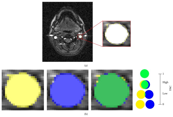
Background and Objectives. The hypothesized link between extracranial venous abnormalities and some neurological disorders awoke interest in the investigation of the internal jugular veins (IJVs). However, different IJV cross-sectional area (CSA) values are currently reported in literature. In this study, we introduced a semiautomatic method to measure and normalize the CSA and the degree of circularity (Circ) of IJVs along their whole length. Methods. Thirty-six healthy subjects (31.22 ± 9.29 years) were recruited and the 2D time-of-flight magnetic resonance venography was acquired with a 1.5 T Siemens scanner. The IJV were segmented on an axial slice, the contours were propagated in 3D. Then, IJV CSA and Circ were computed between the first and the seventh cervical levels (C1-C7) and normalized among subjects. Inter- and intrarater repeatability were assessed. Results. IJV CSA and Circ were significantly different among cervical levels (p < 0.001). A trend for side difference was observed for CSA (larger right IJV, p = 0.06), but not for Circ (p = 0.5). Excellent inter- and intrarater repeatability was obtained for all the measures. Conclusion. This study proposed a reliable semiautomatic method able to measure the IJV area and shape along C1-C7, and suitable for defining the normality thresholds for future clinical studies.
Figures


Similar articles
-
Internal Jugular Vein Cross-Sectional Area Enlargement Is Associated with Aging in Healthy Individuals.PLoS One. 2016 Feb 19;11(2):e0149532. doi: 10.1371/journal.pone.0149532. eCollection 2016. PLoS One. 2016. PMID: 26895434 Free PMC article.
-
Clinical significance of the cross-sectional area of the internal jugular vein.J Cardiothorac Vasc Anesth. 2013 Aug;27(4):685-9. doi: 10.1053/j.jvca.2012.10.007. Epub 2013 Apr 30. J Cardiothorac Vasc Anesth. 2013. PMID: 23642889
-
Magnetic Resonance Imaging of Internal Jugular Veins in Multiple Sclerosis: Interobserver Agreement and Comparison with Doppler Ultrasound Examination.Ann Vasc Surg. 2017 Jul;42:84-92. doi: 10.1016/j.avsg.2016.10.060. Epub 2017 Mar 11. Ann Vasc Surg. 2017. PMID: 28300678
-
Bilateral duplicated internal jugular veins: case study and literature review.Clin Anat. 2007 Apr;20(3):260-6. doi: 10.1002/ca.20366. Clin Anat. 2007. PMID: 16838288 Review.
-
Quantitative flow measurements in the internal jugular veins of multiple sclerosis patients using magnetic resonance imaging.Rev Recent Clin Trials. 2012 May;7(2):117-26. doi: 10.2174/157488712800100206. Rev Recent Clin Trials. 2012. PMID: 22356242 Review.
Cited by
-
Five-Year Longitudinal Study of Neck Vessel Cross-Sectional Area in Multiple Sclerosis.AJNR Am J Neuroradiol. 2018 Sep;39(9):1703-1709. doi: 10.3174/ajnr.A5738. Epub 2018 Jul 26. AJNR Am J Neuroradiol. 2018. PMID: 30049718 Free PMC article.
-
Neck Vessel Cross-Sectional Area Measured with MRI: Scan-Rescan Reproducibility for Longitudinal Evaluations.J Neuroimaging. 2018 Jan;28(1):48-56. doi: 10.1111/jon.12488. Epub 2017 Dec 4. J Neuroimaging. 2018. PMID: 29205670 Free PMC article.
-
Jugular Anomalies in Multiple Sclerosis Are Associated with Increased Collateral Venous Flow.AJNR Am J Neuroradiol. 2017 Aug;38(8):1617-1622. doi: 10.3174/ajnr.A5219. Epub 2017 May 25. AJNR Am J Neuroradiol. 2017. PMID: 28546249 Free PMC article.
-
Ultrasound Evaluation of Internal Jugular Venous Insufficiency and Its Association with Cognitive Decline.Diagnostics (Basel). 2025 Jun 4;15(11):1427. doi: 10.3390/diagnostics15111427. Diagnostics (Basel). 2025. PMID: 40506999 Free PMC article.
-
Connecting the dots: Linking superior ophthalmic vein and internal jugular vein diameter to carotid cavernous fistula type and location.Surg Neurol Int. 2024 Oct 18;15:377. doi: 10.25259/SNI_601_2024. eCollection 2024. Surg Neurol Int. 2024. PMID: 39524569 Free PMC article.
References
MeSH terms
LinkOut - more resources
Full Text Sources
Other Literature Sources
Miscellaneous
