

Nasopharyngeal Tuberculosis: Epidemiology, Mechanism of Infection, Clinical Manifestations, and Management
- PMID: 27034677
- PMCID: PMC4789561
- DOI: 10.1155/2016/4817429
Nasopharyngeal Tuberculosis: Epidemiology, Mechanism of Infection, Clinical Manifestations, and Management
Abstract
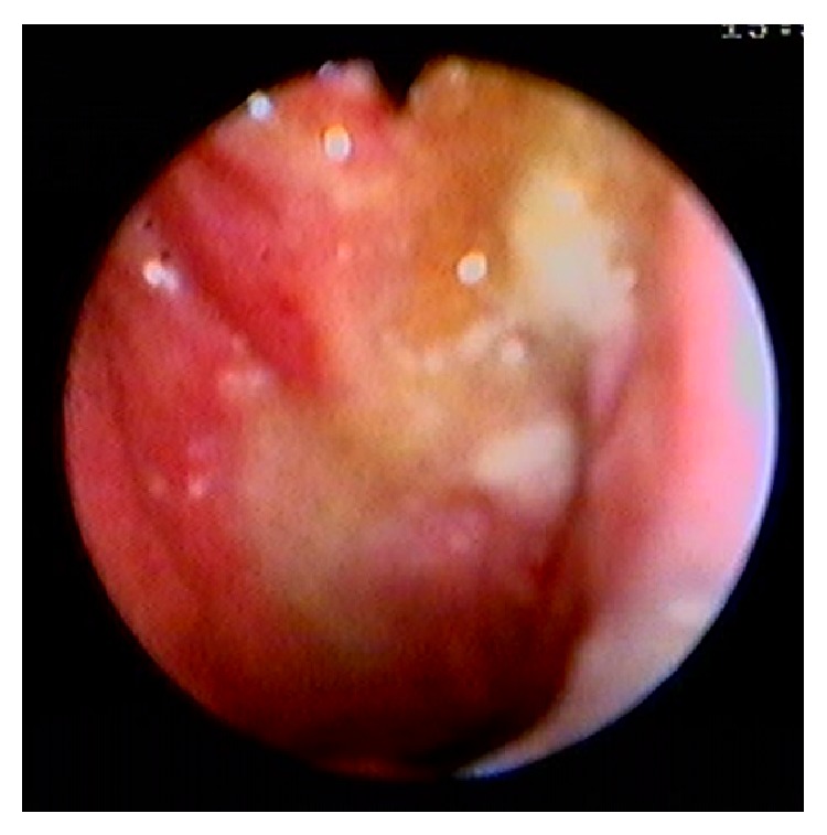
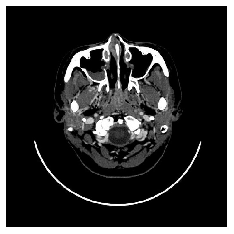
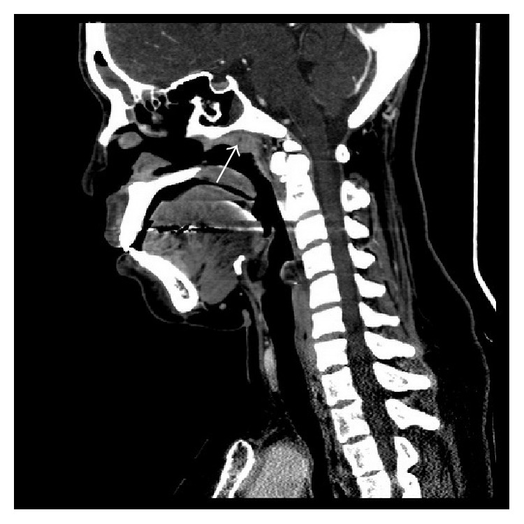
Nasopharyngeal tuberculosis (NPTB) is a noteworthy disease especially in its worldwide spread of the Mycobacterium infection. Although NPTB has been identified in less than one percent of TB cases, recent multiple case reports indicate an either increased awareness or incidence of this disease. The most helpful diagnostic tool is an uncomplicated nasopharyngeal biopsy. However, NPTB is usually ignored because it has varied clinical manifestations and similar presentations with other more common head and neck diseases. Furthermore, the most common presenting symptom is cervical lymphadenopathy mimicking nasopharyngeal carcinoma, a more common and serious disease. Treatment outcomes of NPTB are good in both HIV-positive or HIV-negative patients. In addition, pulmonary tuberculosis association was reported in wide range between 8.3% and 82% which should be considered in a treatment program. In conclusion, early diagnosis and management in NPTB can be achieved by (1) increased awareness of this disease, (2) improvement in knowledge regarding clinical manifestations, and (3) improvement of diagnostic techniques.
Figures
References
-
- WHO. Global Tuberculosis Report. 2014. (WHO Report).
-
- Crown S., Watkins S., Rothholz A. S. Tonsillar and nasopharyngeal infections. Bulletin of the Johns Hopkins Hospital. 1917;28:p. 1.
-
- Thompson S. C. Disease of the Nose and Throat. 2nd. Clasell & CO; 1919.
Publication types
LinkOut - more resources
Full Text Sources
Other Literature Sources
