

Nucleolar PARP-1 Expression Is Decreased in Alzheimer's Disease: Consequences for Epigenetic Regulation of rDNA and Cognition
- PMID: 27034851
- PMCID: PMC4789469
- DOI: 10.1155/2016/8987928
Nucleolar PARP-1 Expression Is Decreased in Alzheimer's Disease: Consequences for Epigenetic Regulation of rDNA and Cognition
Abstract
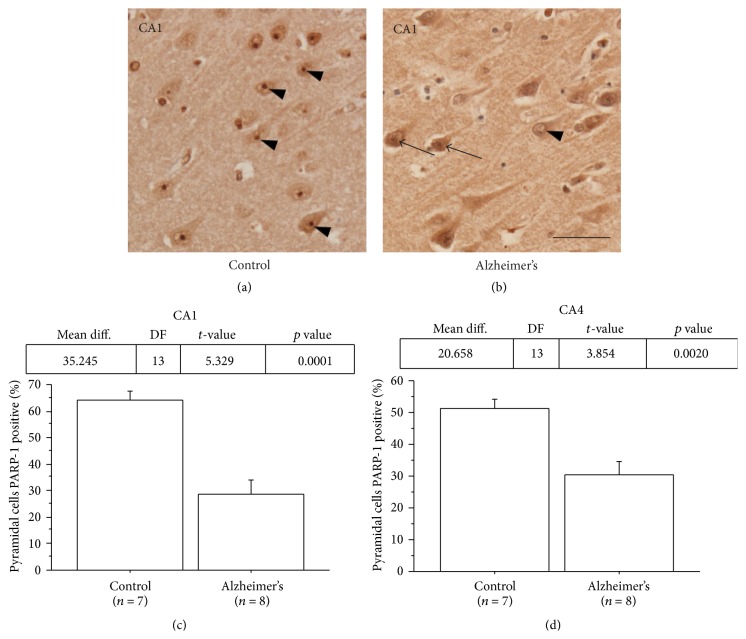
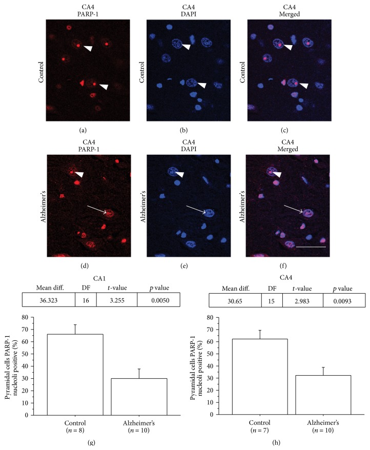
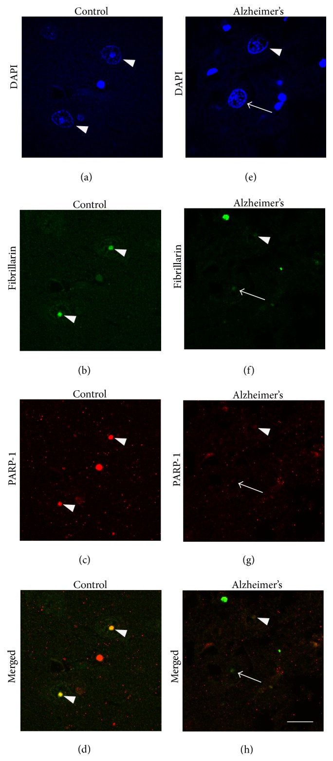
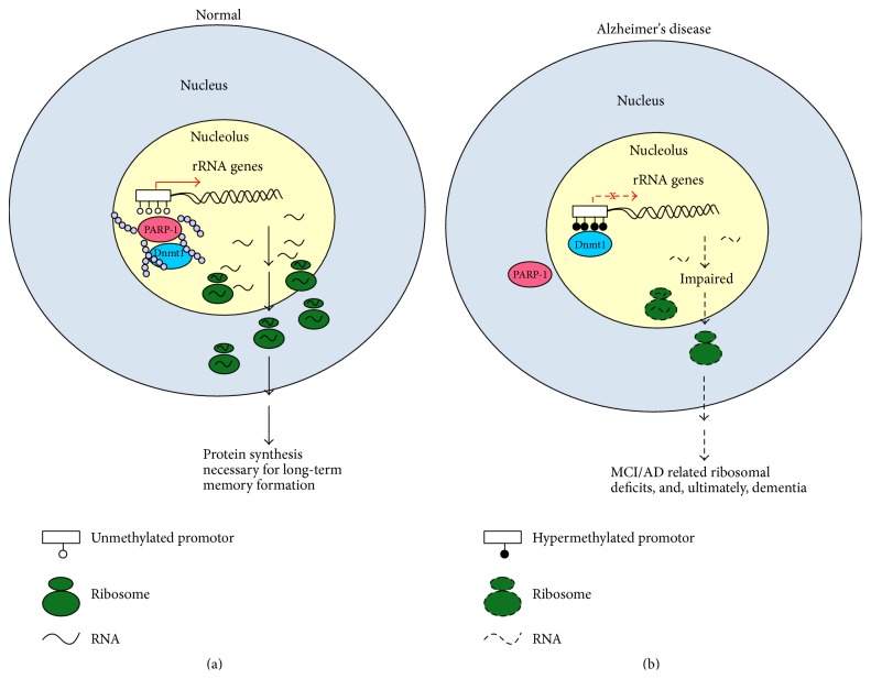
Synaptic dysfunction is thought to play a major role in memory impairment in Alzheimer's disease (AD). PARP-1 has been identified as an epigenetic regulator of plasticity and memory. Thus, we hypothesize that PARP-1 may be altered in postmortem hippocampus of individuals with AD compared to age-matched controls without neurologic disease. We found a reduced level of PARP-1 nucleolar immunohistochemical staining in hippocampal pyramidal cells in AD. Nucleolar PARP-1 staining ranged from dispersed and less intense to entirely absent in AD compared to the distinct nucleolar localization in hippocampal pyramidal neurons in controls. In cases of AD, the percentage of hippocampal pyramidal cells with nucleoli that were positive for both PARP-1 and the nucleolar marker fibrillarin was significantly lower than in controls. PARP-1 nucleolar expression emerges as a sensitive marker of functional changes in AD and suggests a novel role for PARP-1 dysregulation in AD pathology.
Figures
Similar articles
-
Evidence for Decreased Nucleolar PARP-1 as an Early Marker of Cognitive Impairment.Neural Plast. 2019 Nov 19;2019:4383258. doi: 10.1155/2019/4383258. eCollection 2019. Neural Plast. 2019. PMID: 31827497 Free PMC article.
-
PARP-1 and PARP-2 interact with nucleophosmin/B23 and accumulate in transcriptionally active nucleoli.J Cell Sci. 2005 Jan 1;118(Pt 1):211-22. doi: 10.1242/jcs.01606. J Cell Sci. 2005. PMID: 15615785
-
Epigenetic silencing of nucleolar rRNA genes in Alzheimer's disease.PLoS One. 2011;6(7):e22585. doi: 10.1371/journal.pone.0022585. Epub 2011 Jul 22. PLoS One. 2011. PMID: 21799908 Free PMC article.
-
CRTC1 gene is differentially methylated in the human hippocampus in Alzheimer's disease.Alzheimers Res Ther. 2016 Apr 19;8(1):15. doi: 10.1186/s13195-016-0183-0. Alzheimers Res Ther. 2016. PMID: 27094739 Free PMC article.
-
Pyramidal neurone modulation: a therapeutic target for Alzheimer's disease.Neurodegeneration. 1996 Dec;5(4):461-5. doi: 10.1006/neur.1996.0063. Neurodegeneration. 1996. PMID: 9117563 Review.
Cited by
-
The Nucleolus and PARP1 in Cancer Biology.Cancers (Basel). 2020 Jul 6;12(7):1813. doi: 10.3390/cancers12071813. Cancers (Basel). 2020. PMID: 32640701 Free PMC article. Review.
-
Contributions of DNA Damage to Alzheimer's Disease.Int J Mol Sci. 2020 Feb 28;21(5):1666. doi: 10.3390/ijms21051666. Int J Mol Sci. 2020. PMID: 32121304 Free PMC article. Review.
-
Effect of Aflatoxin B1 on the Nervous System: A Systematic Review and Network Analysis Highlighting Alzheimer's Disease.Biology (Basel). 2025 Apr 17;14(4):436. doi: 10.3390/biology14040436. Biology (Basel). 2025. PMID: 40282301 Free PMC article. Review.
-
Polyamine Dysregulation and Nucleolar Disruption in Alzheimer's Disease.J Alzheimers Dis. 2024;98(3):837-857. doi: 10.3233/JAD-231184. J Alzheimers Dis. 2024. PMID: 38489184 Free PMC article.
-
Neuroprotective Effects of PARP Inhibitors in Drosophila Models of Alzheimer's Disease.Cells. 2022 Apr 9;11(8):1284. doi: 10.3390/cells11081284. Cells. 2022. PMID: 35455964 Free PMC article.
References
-
- Masliah E. Mechanisms of synaptic dysfunction in Alzheimer's disease. Histology and Histopathology. 1995;10(2):509–519. - PubMed
Publication types
MeSH terms
Substances
Grants and funding
LinkOut - more resources
Full Text Sources
Other Literature Sources
Medical
Miscellaneous
