. 2016:2016:9576503.
doi: 10.1155/2016/9576503. Epub 2016 Feb 29.

Study of Valproic Acid-Enhanced Hepatocyte Steatosis

Affiliations

Study of Valproic Acid-Enhanced Hepatocyte Steatosis

Renin Chang et al. Biomed Res Int. 2016.

Abstract

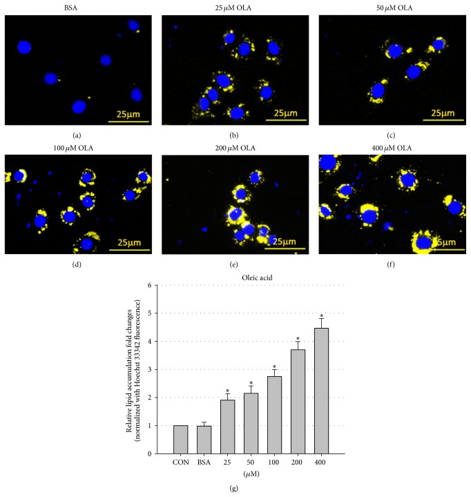
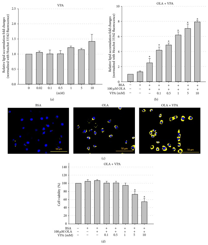
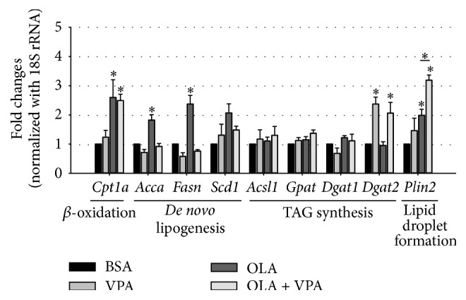
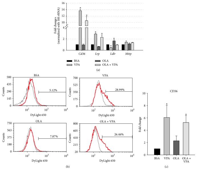
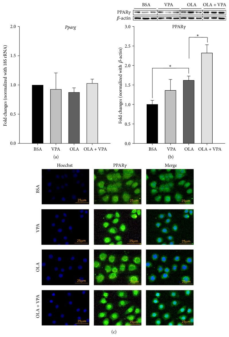
Valproic acid (VPA) is one of the most widely used antiepilepsy drugs. However, several side effects, including weight gain and fatty liver, have been reported in patients following VPA treatment. In this study, we explored the molecular mechanisms of VPA-induced hepatic steatosis using FL83B cell line-based in vitro model. Using fluorescent lipid staining technique, we found that VPA enhanced oleic acid- (OLA-) induced lipid accumulation in a dose-dependent manner in hepatocytes; this may be due to upregulated lipid uptake, triacylglycerol (TAG) synthesis, and lipid droplet formation. Real-time PCR results showed that, following VPA treatment, the expression levels of genes encoding cluster of differentiation 36 (Cd36), low-density lipoprotein receptor-related protein 1 (Lrp1), diacylglycerol acyltransferase 2 (Dgat2), and perilipin 2 (Plin2) were increased, that of carnitine palmitoyltransferase I a (Cpt1a) was not affected, and those of acetyl-Co A carboxylase α (Acca) and fatty acid synthase (Fasn) were decreased. Furthermore, using immunofluorescence staining and flow cytometry analyses, we found that VPA also induced peroxisome proliferator-activated receptor γ (PPARγ) nuclear translocation and increased levels of cell-surface CD36. Based on these results, we propose that VPA may enhance OLA-induced hepatocyte steatosis through the upregulation of PPARγ- and CD36-dependent lipid uptake, TAG synthesis, and lipid droplet formation.


Figures

Figure 1
Figure 1
OLA induces lipid accumulation in FL83B cells in a dose-dependent manner. Microscopic images of FL83B cells treated with BSA only (a), or 25 μM (b), 50 μM (c), 100 μM (d), 200 μM (e), and 400 μM (f) BSA-conjugated OLA for 24 hours. Cells were fixed and stained with Hoechst 33342 (blue, nucleus) and Nile Red (yellow, intracellular lipid droplets). Quantification of intracellular lipid accumulation was performed using a fluorescent microplate reader (g). Values are means ± SEM of four independent experiments. above the bars refer to significant differences (P < 0.05). CON: control, treated with normal culture medium only.
Figure 2
Figure 2
VPA enhances OLA-induced lipid accumulation in FL83B cells and induces cytotoxicity. Nile Red and Hoechst 33342 double staining showed that 24-hour VPA single treatment (a) did not significantly induce steatosis but enhanced OLA- (100 μM) induced intracellular neutral lipid accumulation (b) and lipid droplet formation (OLA = 100 μM, VPA = 1 mM) (c) in a dose-dependent manner in FL83B cells. MTT cell viability results also indicate high doses (5 and 10 mM) of VPA-induced lipotoxicity in FL83B cells (d). Values are means ± SEM of four independent experiments. above the bars refer to significant differences (P < 0.05).
Figure 3
Figure 3
Regulation of expression of lipid metabolism-related genes by OLA and VPA. The mRNA levels of lipid catabolism/anabolism-related genes including Cpt1, Acca, Fasn, Scd1, Acsl1, Gpat, Dgat1, Dgat2, and Plin2 in FL83B cells treated with BSA, 100 μM OLA, 1 mM VPA, or 100 μM OLA plus 1 mM VPA for 24 hours were analyzed using real-time PCR. Relative fold changes were calculated using Ct values obtained from three independent experiments and are shown as means ± SEM. above the bars refer to significant differences (P < 0.05).
Figure 4
Figure 4
VPA increases the mRNA and cell-surface protein expression levels of fatty acid translocase CD36. The mRNA expression levels of lipid transport-related genes including Cd36, Lrp, Ldlr, and Mttp were analyzed using real-time PCR (a). Relative fold changes were calculated using Ct values obtained from three independent experiments and are shown as means ± SEM. above the bars refer to significant differences (P < 0.05). The Fl83B cell-surface CD36 expression levels were analyzed by flow cytometry using mouse anti-CD36 antibody and DyLight 650-conjugated secondary antibody, and representative histograms (b) and quantitative flow cytometry data (c) are shown. above the bars refer to significant differences (P < 0.05). For background fluorescent subtraction, mouse isotype IgG antibody was used (OLA = 100 μM, VPA = 1 mM, 24 hours).
Figure 5
Figure 5
VPA enhances oleic acids increased PPARγ protein expression and nuclear translocation, but not the mRNA levels. Real-time PCR (a), Western blotting (b), and immunofluorescence staining (c) were conducted following 24-hour treatment with BSA, VPA (1 mM), OLA (100 μM), or OLA plus VPA. For real-time PCR, relative fold changes were calculated using Ct values obtained from three independent experiments and are shown as means ± SEM. above the bars refer to significant differences (P < 0.05). Densitometric analyses for Western blotting were conducted for sample sets obtained from three independent experiments, and results are shown as means ± SEM. above the bars refer to significant differences (P < 0.05). In the immunofluorescence staining images, nuclear and intracellular PPARγ proteins were stained by Hoechst 33342- (blue) and DyLight 488-conjugated antibody (green), respectively.

References

    1. Saleh D. A. A., Ismail M. A., Ibrahim A. M. Non alcoholic fatty liver disease, insulin resistance, dyslipidemia and atherogenic ratios in epileptic children and adolescents on long term antiepileptic drug therapy. Pakistan Journal of Biological Sciences. 2012;15(2):68–77. doi: 10.3923/pjbs.2012.68.77. - DOI - PubMed
    1. Luef G. J., Waldmann M., Sturm W., et al. Valproate therapy and nonalcoholic fatty liver disease. Annals of Neurology. 2004;55(5):729–732. doi: 10.1002/ana.20074. - DOI - PubMed
    1. Verrotti A., Di Marco G., La Torre R., Pelliccia P., Chiarelli F. Nonalcoholic fatty liver disease during valproate therapy. European Journal of Pediatrics. 2009;168(11):1391–1394. doi: 10.1007/s00431-009-0927-y. - DOI - PubMed
    1. Knapp A. C., Todesco L., Beier K., et al. Toxicity of valproic acid in mice with decreased plasma and tissue carnitine stores. Journal of Pharmacology and Experimental Therapeutics. 2008;324(2):568–575. doi: 10.1124/jpet.107.131185. - DOI - PubMed
    1. Peterson G. M., Naunton M. Valproate: a simple chemical with so much to offer. Journal of Clinical Pharmacy and Therapeutics. 2005;30(5):417–421. doi: 10.1111/j.1365-2710.2005.00671.x. - DOI - PubMed

Publication types

MeSH terms

LinkOut - more resources