

Upregulated ATF6 contributes to chronic intermittent hypoxia-afforded protection against myocardial ischemia/reperfusion injury
- PMID: 27035093
- PMCID: PMC4829135
- DOI: 10.3892/ijmm.2016.2535
Upregulated ATF6 contributes to chronic intermittent hypoxia-afforded protection against myocardial ischemia/reperfusion injury
Abstract
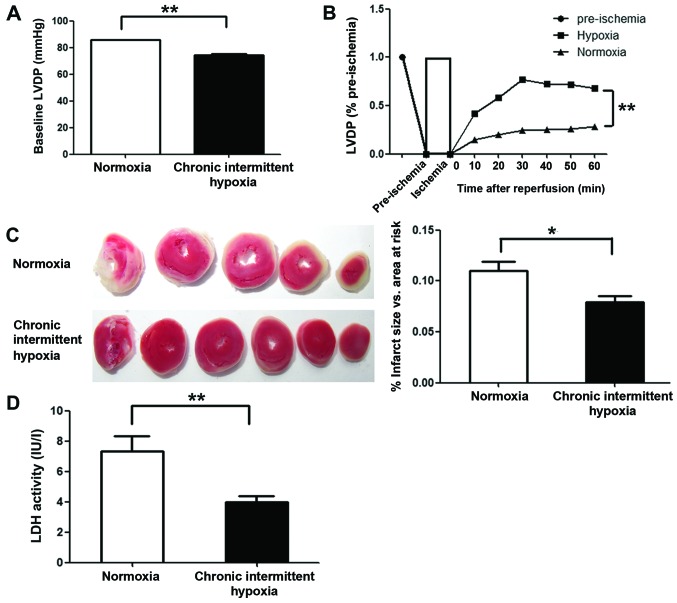
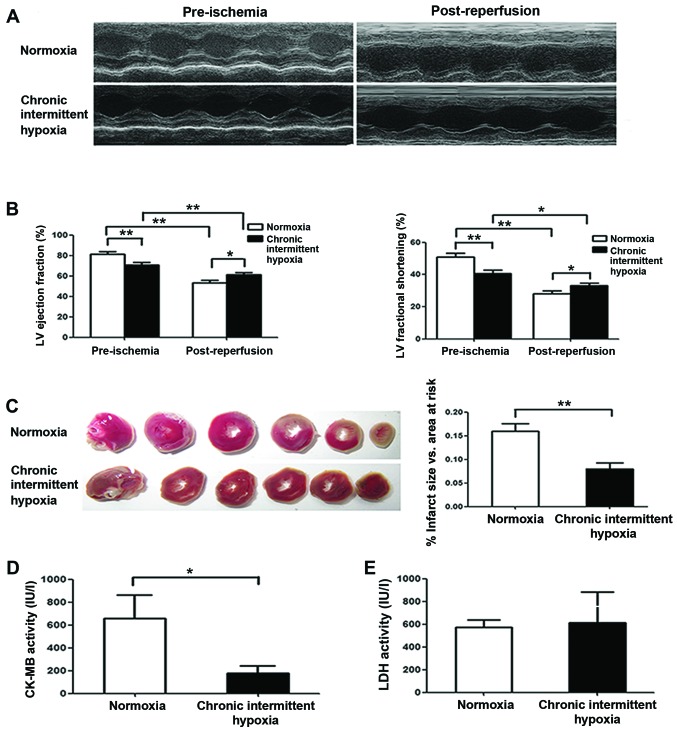
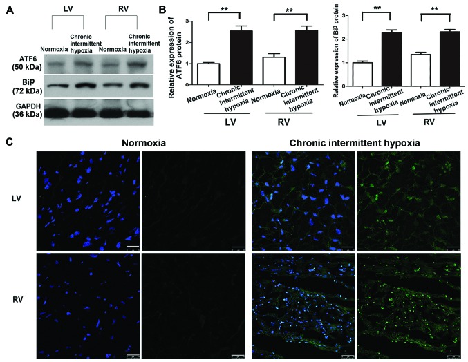
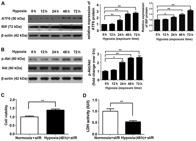
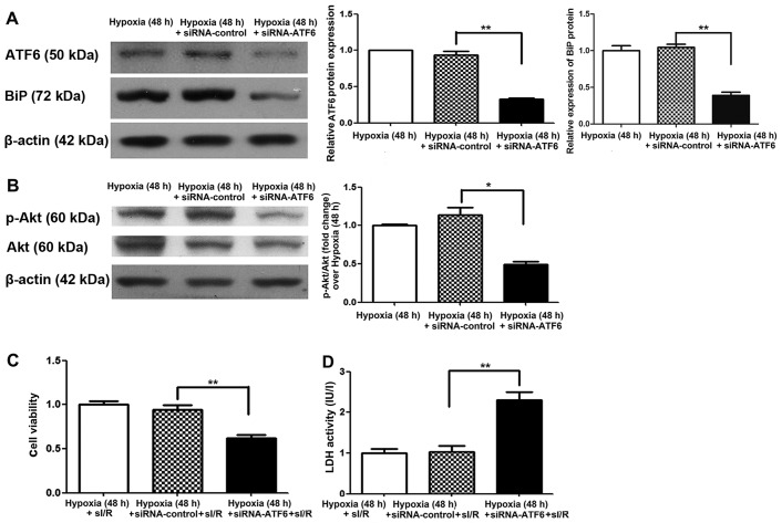
In the present study, we investigated the role of activating transcription factor 6 (ATF6) in the mechanism by which chronic intermittent hypoxia (CIH) increases tolerance to myocardial ischemia/reperfusion (I/R). Experiments were conducted using a rat model of I/R injury in vivo and isolated Langendorff-perfused rat hearts ex vivo. The role of Akt in this process was also investigated in vitro using rat myoblast H9c2 cells. Cell viability was measured using a cell counting kit-8 assay. Lactate dehydrogenase (LDH) and creatine kinase cardiac isoenzyme activity were also measured as markers of cellular damage. ATF6, Akt and phosphorylated (p)-Akt expression was analyzed by western blot analysis. RNA interference (RNAi) was used to suppress ATF6 expression. We noted that ATF6 expression in the ventricular myocardium was significantly increased in rats exposed to CIH. Furthermore, we noted that CIH preserved cardiac function after I/R in vivo and improved post-ischemic recovery of myocardial performance in isolated rat hearts. ATF6 and p-Akt expression was upregulated in cultured H9c2 cells exposed to chronic mild hypoxia compared with those cultured under normoxic conditions. Chronic mild hypoxia attenuated subsequent simulated I/R injury in H9c2 cells (48 h), as evidenced by increased cell viability and decreased LDH activity. By contrast, decreased cell viability and increased LDH activity were observed in siRNA-ATF6-transfected H9c2 cells, with a concomitant reduction in p-Akt levels. These results indicated that ATF6 upregulation is involved in the mechanism by which CIH attenuates myocardial I/R injury, possibly through upregulation of p-Akt, which is a key regulator of cardiomyocyte survival.
Figures
Similar articles
-
Trimetazidine protects against myocardial ischemia/reperfusion injury by inhibiting excessive autophagy.J Mol Med (Berl). 2018 Aug;96(8):791-806. doi: 10.1007/s00109-018-1664-3. Epub 2018 Jun 29. J Mol Med (Berl). 2018. PMID: 29955901
-
ROS generated during early reperfusion contribute to intermittent hypobaric hypoxia-afforded cardioprotection against postischemia-induced Ca(2+) overload and contractile dysfunction via the JAK2/STAT3 pathway.J Mol Cell Cardiol. 2015 Apr;81:150-61. doi: 10.1016/j.yjmcc.2015.02.015. Epub 2015 Feb 27. J Mol Cell Cardiol. 2015. PMID: 25731682
-
Remifentanil Preconditioning Reduces Postischemic Myocardial Infarction and Improves Left Ventricular Performance via Activation of the Janus Activated Kinase-2/Signal Transducers and Activators of Transcription-3 Signal Pathway and Subsequent Inhibition of Glycogen Synthase Kinase-3β in Rats.Crit Care Med. 2016 Mar;44(3):e131-45. doi: 10.1097/CCM.0000000000001350. Crit Care Med. 2016. PMID: 26468894
-
Magnesium lithospermate B improves myocardial function and prevents simulated ischemia/reperfusion injury-induced H9c2 cardiomyocytes apoptosis through Akt-dependent pathway.J Ethnopharmacol. 2014;151(1):714-21. doi: 10.1016/j.jep.2013.11.036. Epub 2013 Dec 1. J Ethnopharmacol. 2014. PMID: 24296090
-
Ginsenoside Rg3 attenuates myocardial ischemia/reperfusion injury via Akt/endothelial nitric oxide synthase signaling and the B‑cell lymphoma/B‑cell lymphoma‑associated X protein pathway.Mol Med Rep. 2015 Jun;11(6):4518-24. doi: 10.3892/mmr.2015.3336. Epub 2015 Feb 11. Mol Med Rep. 2015. PMID: 25672441
Cited by
-
Intermittent Hypoxia Prevents Myocardial Mitochondrial Ca2+ Overload and Cell Death during Ischemia/Reperfusion: The Role of Reactive Oxygen Species.Cells. 2019 Jun 9;8(6):564. doi: 10.3390/cells8060564. Cells. 2019. PMID: 31181855 Free PMC article.
-
Protein homeostasis in the aged and diseased heart.J Cardiovasc Aging. 2023;3(2):16. doi: 10.20517/jca.2023.4. Epub 2023 Mar 7. J Cardiovasc Aging. 2023. PMID: 37092014 Free PMC article.
-
Ubiquinol-cytochrome c reductase core protein 1 contributes to cardiac tolerance to acute exhaustive exercise.Exp Biol Med (Maywood). 2022 Jan;247(2):165-173. doi: 10.1177/15353702211046546. Epub 2021 Oct 14. Exp Biol Med (Maywood). 2022. PMID: 34648372 Free PMC article.
-
Network pharmacology-based identification of major component of Angelica sinensis and its action mechanism for the treatment of acute myocardial infarction.Biosci Rep. 2018 Nov 7;38(6):BSR20180519. doi: 10.1042/BSR20180519. Print 2018 Dec 21. Biosci Rep. 2018. PMID: 30232231 Free PMC article.
-
Unfolded protein response (UPR) integrated signaling networks determine cell fate during hypoxia.Cell Mol Biol Lett. 2020 Mar 13;25:18. doi: 10.1186/s11658-020-00212-1. eCollection 2020. Cell Mol Biol Lett. 2020. PMID: 32190062 Free PMC article. Review.
References
Publication types
MeSH terms
Substances
LinkOut - more resources
Full Text Sources
Other Literature Sources
