

Nemo-like kinase regulates the expression of vascular endothelial growth factor (VEGF) in alveolar epithelial cells
- PMID: 27035511
- PMCID: PMC4817507
- DOI: 10.1038/srep23987
Nemo-like kinase regulates the expression of vascular endothelial growth factor (VEGF) in alveolar epithelial cells
Abstract
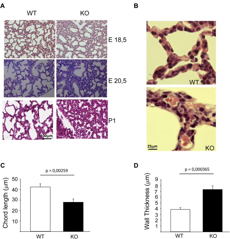
The canonical Wnt signaling can be silenced either through β-catenin-mediated ubiquitination and degradation or through phosphorylation of Tcf and Lef by nemo-like kinase (NLK). In the present study, we generated NLK deficient animals and found that these mice become cyanotic shortly before death because of lung maturation defects. NLK-/- lungs exhibited smaller and compressed alveoli and the mesenchyme remained thick and hyperplastic. This phenotype was caused by epithelial activation of vascular endothelial growth factor (VEGF) via recruitment of Lef1 to the promoter of VEGF. Elevated expression of VEGF and activation of the VEGF receptor through phosphorylation promoted an increase in the proliferation rate of epithelial and endothelial cells. In summary, our study identifies NLK as a novel signaling molecule for proper lung development through the interconnection between epithelial and endothelial cells during lung morphogenesis.
Figures


Similar articles
-
NLK-mediated phosphorylation of HDAC1 negatively regulates Wnt signaling.Mol Biol Cell. 2017 Jan 15;28(2):346-355. doi: 10.1091/mbc.E16-07-0547. Epub 2016 Nov 30. Mol Biol Cell. 2017. PMID: 27903773 Free PMC article.
-
The TAK1-NLK-MAPK-related pathway antagonizes signalling between beta-catenin and transcription factor TCF.Nature. 1999 Jun 24;399(6738):798-802. doi: 10.1038/21674. Nature. 1999. PMID: 10391247
-
Deletion of Nemo-like Kinase in T Cells Reduces Single-Positive CD8+ Thymocyte Population.J Immunol. 2020 Oct 1;205(7):1830-1841. doi: 10.4049/jimmunol.2000109. Epub 2020 Aug 24. J Immunol. 2020. PMID: 32839237
-
Nemo-like kinase, a multifaceted cell signaling regulator.Cell Signal. 2013 Jan;25(1):190-7. doi: 10.1016/j.cellsig.2012.09.017. Epub 2012 Sep 18. Cell Signal. 2013. PMID: 23000342 Review.
-
The emerging role of Nemo-like kinase (NLK) in the regulation of cancers.Tumour Biol. 2015 Dec;36(12):9147-52. doi: 10.1007/s13277-015-4159-7. Epub 2015 Oct 1. Tumour Biol. 2015. PMID: 26427665 Review.
Cited by
-
WNT signaling - lung cancer is no exception.Respir Res. 2017 Sep 5;18(1):167. doi: 10.1186/s12931-017-0650-6. Respir Res. 2017. PMID: 28870231 Free PMC article. Review.
-
Epstein-Barr virus-encoded microRNAs as regulators in host immune responses.Int J Biol Sci. 2018 Apr 5;14(5):565-576. doi: 10.7150/ijbs.24562. eCollection 2018. Int J Biol Sci. 2018. PMID: 29805308 Free PMC article. Review.
-
NLK-mediated phosphorylation of HDAC1 negatively regulates Wnt signaling.Mol Biol Cell. 2017 Jan 15;28(2):346-355. doi: 10.1091/mbc.E16-07-0547. Epub 2016 Nov 30. Mol Biol Cell. 2017. PMID: 27903773 Free PMC article.
-
Nemo-Like Kinase in Development and Diseases: Insights from Mouse Studies.Int J Mol Sci. 2020 Dec 2;21(23):9203. doi: 10.3390/ijms21239203. Int J Mol Sci. 2020. PMID: 33276680 Free PMC article. Review.
-
Diamond Blackfan anemia is mediated by hyperactive Nemo-like kinase.Nat Commun. 2020 Jul 3;11(1):3344. doi: 10.1038/s41467-020-17100-z. Nat Commun. 2020. PMID: 32620751 Free PMC article.
References
-
- Perl A. K. & Whitsett J. A. Molecular mechanisms controlling lung morphogenesis. Clin Genet 56, 14–27 (1999). - PubMed
-
- Korfhagen T. R., Glasser S. W. & Stripp B. R. Regulation of gene expression in the lung. Curr Opin Pediatr. 6, 255–261 (1994). - PubMed
-
- Kumar V. H., Lakshminrusimha S., El Abiad M. T., Chess P. R. & Ryan R. M. Growth factors in lung development. Adv Clin Chem. 40, 261–316 (2005). - PubMed
Publication types
MeSH terms
Substances
LinkOut - more resources
Full Text Sources
Other Literature Sources
Molecular Biology Databases
Miscellaneous
