

Toward a better understanding of the cellular basis for cerebrospinal fluid shunt obstruction: report on the construction of a bank of explanted hydrocephalus devices
- PMID: 27035548
- PMCID: PMC5915300
- DOI: 10.3171/2016.2.PEDS15531
Toward a better understanding of the cellular basis for cerebrospinal fluid shunt obstruction: report on the construction of a bank of explanted hydrocephalus devices
Abstract
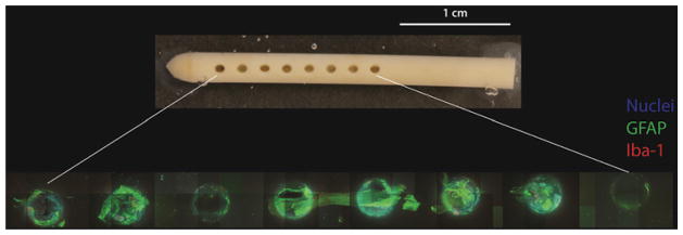
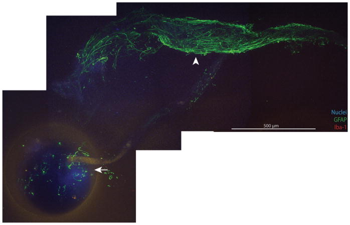
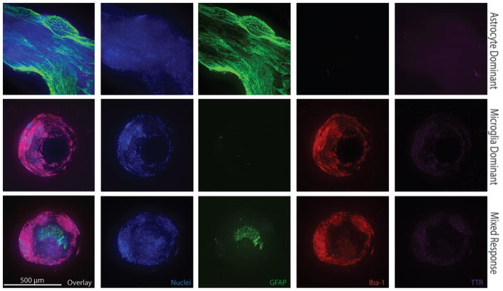
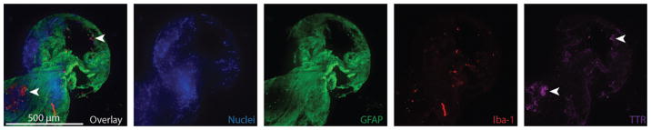
OBJECTIVE Shunt obstruction by cells and/or tissue is the most common cause of shunt failure. Ventricular catheter obstruction alone accounts for more than 50% of shunt failures in pediatric patients. The authors sought to systematically collect explanted ventricular catheters from the Seattle Children's Hospital with a focus on elucidating the cellular mechanisms underlying obstruction. METHODS In the operating room, explanted hardware was placed in 4% paraformaldehyde. Weekly, samples were transferred to buffer solution and stored at 4°C. After consent was obtained for their use, catheters were labeled using cell-specific markers for astrocytes (glial fibrillary acidic protein), microglia (ionized calcium-binding adapter molecule 1), and choroid plexus (transthyretin) in conjunction with a nuclear stain (Hoechst). Catheters were mounted in custom polycarbonate imaging chambers. Three-dimensional, multispectral, spinning-disk confocal microscopy was used to image catheter cerebrospinal fluid-intake holes (10× objective, 499.2-μm-thick z-stack, 2.4-μm step size, Olympus IX81 inverted microscope with motorized stage and charge-coupled device camera). Values are reported as the mean ± standard error of the mean and were compared using a 2-tailed Mann-Whitney U-test. Significance was defined at p < 0.05. RESULTS Thirty-six ventricular catheters have been imaged to date, resulting in the following observations: 1) Astrocytes and microglia are the dominant cell types bound directly to catheter surfaces; 2) cellular binding to catheters is ubiquitous even if no grossly visible tissue is apparent; and 3) immunohistochemical techniques are of limited utility when a catheter has been exposed to Bugbee wire electrocautery. Statistical analysis of 24 catheters was performed, after excluding 7 catheters exposed to Bugbee wire cautery, 3 that were poorly fixed, and 2 that demonstrated pronounced autofluorescence. This analysis revealed that catheters with a microglia-dominant cellular response tended to be implanted for shorter durations (24.7 ± 6.7 days) than those with an astrocyte-dominant response (1183 ± 642 days; p = 0.027). CONCLUSIONS Ventricular catheter occlusion remains a significant source of shunt morbidity in the pediatric population, and given their ability to intimately associate with catheter surfaces, astrocytes and microglia appear to be critical to this pathophysiology. Microglia tend to be the dominant cell type on catheters implanted for less than 2 months, while astrocytes tend to be the most prevalent cell type on catheters implanted for longer time courses and are noted to serve as an interface for the secondary attachment of ependymal cells and choroid plexus.
Keywords: CSF = cerebrospinal fluid; GFAP = glial fibrillary acidic protein; HBHS = HEPES-buffered Hanks solution; HIPAA = Health Insurance Portability and Accountability Act; IHC = immunohistochemical; IRB = institutional review board; PDMS = poly(dimethylsiloxane); PFA = paraformaldehyde; RT = room temperature; SCH = Seattle Children's Hospital; TTR = transthyretin; astrocyte; hydrocephalus; microglia; shunt failure; shunt obstruction; ventriculoperitoneal shunt.
Figures

References
-
- Aschoff A, Kremer P, Hashemi B, Kunze S. The scientific history of hydrocephalus and its treatment. Neurosurg Rev. 1999;22:67–95. - PubMed
-
- Baru JS, Bloom DA, Muraszko K, Koop CE. John Holter’s shunt. J Am Coll Surg. 2001;192:79–85. - PubMed
-
- Bondurant CP, Jimenez DF. Epidemiology of cerebrospinal fluid shunting. Pediatr Neurosurg. 1995;23:254–259. - PubMed
-
- Browd SR, Ragel BT, Gottfried ON, Kestle JR. Failure of cerebrospinal fluid shunts: part I: Obstruction and mechanical failure. Pediatr Neurol. 2006;34:83–92. - PubMed
-
- Davalos D, Grutzendler J, Yang G, Kim JV, Zuo Y, Jung S, et al. ATP mediates rapid microglial response to local brain injury in vivo. Nat Neurosci. 2005;8:752–758. - PubMed
MeSH terms
Grants and funding
LinkOut - more resources
Full Text Sources
Other Literature Sources
Medical
Molecular Biology Databases
Research Materials
Miscellaneous
