Review
. 2016 Jun;37(3):190-222.
doi: 10.1210/er.2015-1116. Epub 2016 Apr 1.

Monogenic Diabetes: What It Teaches Us on the Common Forms of Type 1 and Type 2 Diabetes

Affiliations
Review

Monogenic Diabetes: What It Teaches Us on the Common Forms of Type 1 and Type 2 Diabetes

Yisheng Yang et al. Endocr Rev. 2016 Jun.

Abstract

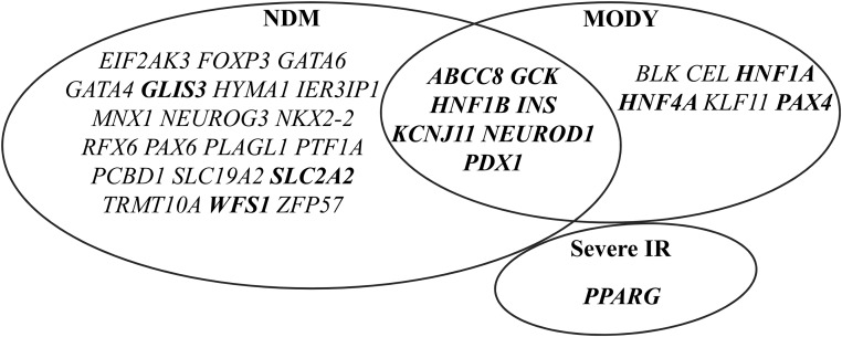
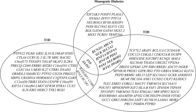
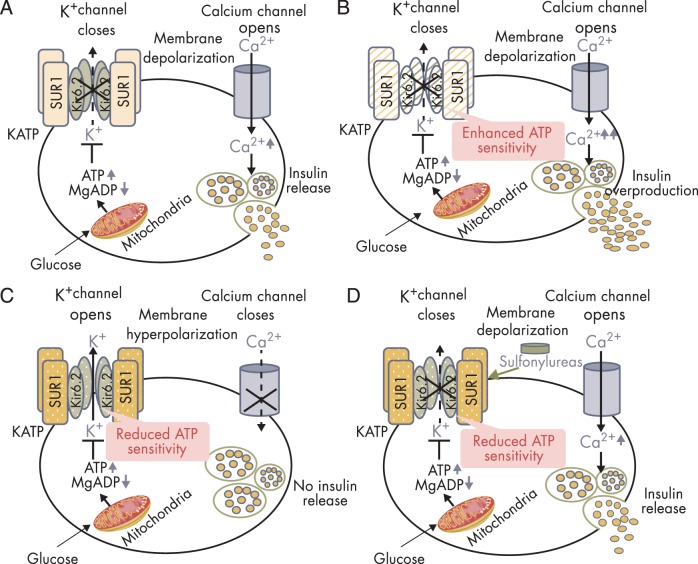
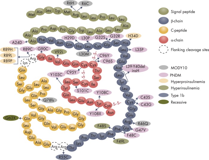
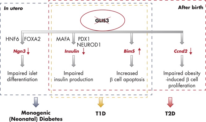
To date, more than 30 genes have been linked to monogenic diabetes. Candidate gene and genome-wide association studies have identified > 50 susceptibility loci for common type 1 diabetes (T1D) and approximately 100 susceptibility loci for type 2 diabetes (T2D). About 1-5% of all cases of diabetes result from single-gene mutations and are called monogenic diabetes. Here, we review the pathophysiological basis of the role of monogenic diabetes genes that have also been found to be associated with common T1D and/or T2D. Variants of approximately one-third of monogenic diabetes genes are associated with T2D, but not T1D. Two of the T2D-associated monogenic diabetes genes-potassium inward-rectifying channel, subfamily J, member 11 (KCNJ11), which controls glucose-stimulated insulin secretion in the β-cell; and peroxisome proliferator-activated receptor γ (PPARG), which impacts multiple tissue targets in relation to inflammation and insulin sensitivity-have been developed as major antidiabetic drug targets. Another monogenic diabetes gene, the preproinsulin gene (INS), is unique in that INS mutations can cause hyperinsulinemia, hyperproinsulinemia, neonatal diabetes mellitus, one type of maturity-onset diabetes of the young (MODY10), and autoantibody-negative T1D. Dominant heterozygous INS mutations are the second most common cause of permanent neonatal diabetes. Moreover, INS gene variants are strongly associated with common T1D (type 1a), but inconsistently with T2D. Variants of the monogenic diabetes gene Gli-similar 3 (GLIS3) are associated with both T1D and T2D. GLIS3 is a key transcription factor in insulin production and β-cell differentiation during embryonic development, which perturbation forms the basis of monogenic diabetes as well as its association with T1D. GLIS3 is also required for compensatory β-cell proliferation in adults; impairment of this function predisposes to T2D. Thus, monogenic forms of diabetes are invaluable "human models" that have contributed to our understanding of the pathophysiological basis of common T1D and T2D.


Figures

Figure 1.
Figure 1.
Monogenic diabetes genes associated with NDM and/or MODY. Over 30 monogenic diabetes genes have been identified to cause NDM or MODY. Mutations in seven of them, including ABCC8, GCK, HNF1B, INS, KCNJ11, NEUROD1, and PDX1, may lead to both NDM and MODY. Bolded genes are discussed in this article.
Figure 2.
Figure 2.
Venn diagram of intersection between the loci/genes associated with T1D or T2D and known monogenic diabetes. Over 50 susceptibility loci for common T1D and approximately 100 susceptibility loci for T2D have been identified by GWAS and candidate gene association studies. INS, GLIS3, RASGRP, COBL, RNLS, and BCAR1 are associated with both T1D and T2D. About one-third of the known monogenic diabetes genes are associated with T2D. INS and GLIS3 are the two known monogenic diabetes genes whose variants are associated with both T1D and T2D.
Figure 3.
Figure 3.
Insulin secretion in normal and KATP channel mutant pancreatic β-cells. A, Glucose-stimulated insulin secretion in normal β-cells. The KATP channel is composed of four Kir6.2 subunits encoded by KCNJ11 and four SUR1 subunits encoded by ABCC8. High glucose in the circulation leads to increased glucose uptake into pancreatic β-cells. Increased intracellular glucose is metabolized via glycolytic and mitochondrial metabolism, leading to an increase in ATP production and a fall in MgADP. This results in KATP channel closure, membrane depolarization, opening of voltage-gated Ca2+ channels, Ca2+ influx, and insulin release. B, Insulin overproduction in the β-cell with KATP channel-inactivating mutations. Loss-of-function mutations in KATP channel enhances ATP binding to the channel, leading to KATP channel closure, membrane depolarization, insulin oversecretion and CHI. C, Impaired insulin secretion in the β-cell with KATP channel activating mutations. Gain-of-function mutations in KATP channel impair ATP binding to the channel Kir6.2 mutant (shown in olive green, encoded by KCNJ11), or enhance the binding of Mg-nucleotide to SUR1 mutant (shown in amber, encoded by ABCC8) leading to KATP channel opening, membrane hyperpolarization, impaired insulin release, and NDM. D, Oral sulfonylureas stimulate insulin secretion in patients with KATP channel-activating mutations. Sulfonylureas bind to the ABCC8-encoded SUR1 subunit of the KATP channel to effect channel closure independent of ATP and enable insulin secretion.
Figure 4.
Figure 4.
Diagram depicting the human preproinsulin molecule with mutations that are currently known to cause diabetes and hyperinsulinemia or hyperproinsulinemia. The amino acid residues in the signal peptide, the β-chain, the C-peptide, and the α-chain are shown in different colors. The dashed circles indicate the basic residues that are the cleavage sites for conversion from proinsulin to insulin. The mutations underlying different types of diabetes and hyperinsulinemia or hyperproinsulinemia are presented in oval shapes filled in distinct colors as indicated. We note that some translation initiation codon mutations (eg, 3G>T, 3G>A), in-frame deletion mutants in the coding region, and mutations in the promoter or intron regions of the INS gene have been left out of this diagram. In addition, several INS substitution mutants, eg, A23S, A23T, L68M, and G84R, that were initially reported to occur in patients with diabetes have been left out of this diagram because subsequent studies showed that they are functional incidental variants. (See Section III.A for details.)
Figure 5.
Figure 5.
Multifaceted role of GLIS3 in diabetes. In utero, GLIS3 controls islet differentiation by transactivating Ngn3, synergistically with HNF6 and forkhead box A2 (FOXA2, also known as HNF3B or transcription factor 3B [TCF-3B]). Loss-of-function mutations or ablation of Glis3 cause impaired islet differentiation and NDM. After birth, GLIS3 predominantly controls insulin gene transcription, cooperating with MAFA, PDX1, and NEUROD1. GLIS3 is also required for obesity-induced β-cell proliferation and compensatory β-cell mass expansion by transactivating Ccnd2. In addition, GLIS3 plays a protective role for β-cell survival, possibly by regulating BimS. Therefore, impairment of GLIS3 function also plays a key role in the development of T1D and T2D. This figure summarizes the pathways by which ablation, loss-of-function mutations, or functional impairment of GLIS3 cause NDM, T1D, and T2D. Pathways in the blue rectangle take place in the developing pancreas in utero and underlie monogenic NDM; pathways in the yellow square and the red rectangle pertain to the postnatal β-cell and contribute to the development of T1D and T2D, respectively.

Similar articles

Cited by

References

    1. Steck AK, Winter WE. Review on monogenic diabetes. Curr Opin Endocrinol Diabetes Obes. 2011;18(4):252–258. - PubMed
    1. Greeley SA, Naylor RN, Philipson LH, Bell GI. Neonatal diabetes: an expanding list of genes allows for improved diagnosis and treatment. Curr Diab Rep. 2011;11(6):519–532. - PMC - PubMed
    1. Edghill EL, Dix RJ, Flanagan SE, et al. HLA genotyping supports a nonautoimmune etiology in patients diagnosed with diabetes under the age of 6 months. Diabetes. 2006;55(6):1895–1898. - PubMed
    1. Flanagan SE, Haapaniemi E, Russell MA, et al. Activating germline mutations in STAT3 cause early-onset multi-organ autoimmune disease. Nat Genet. 2014;46(8):812–814. - PMC - PubMed
    1. Fajans SS, Bell GI. MODY: history, genetics, pathophysiology, and clinical decision making. Diabetes Care. 2011;34(8):1878–1884. - PMC - PubMed

Publication types

MeSH terms