

Inactivation of the miR-183/96/182 Cluster Decreases the Severity of Pseudomonas aeruginosa-Induced Keratitis
- PMID: 27035623
- PMCID: PMC4819431
- DOI: 10.1167/iovs.16-19134
Inactivation of the miR-183/96/182 Cluster Decreases the Severity of Pseudomonas aeruginosa-Induced Keratitis
Abstract
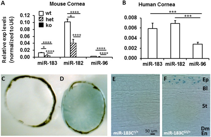
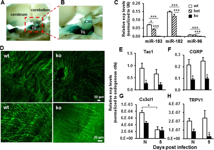
Purpose: The microRNA-183/96/182 cluster (miR-183/96/182) plays important roles in sensory organs. Because the cornea is replete with sensory innervation, we hypothesized that miR-183/96/182 modulates the corneal response to bacterial infection through regulation of neuroimmune interactions.
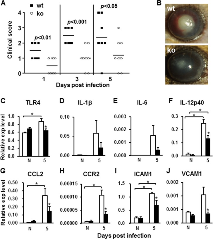
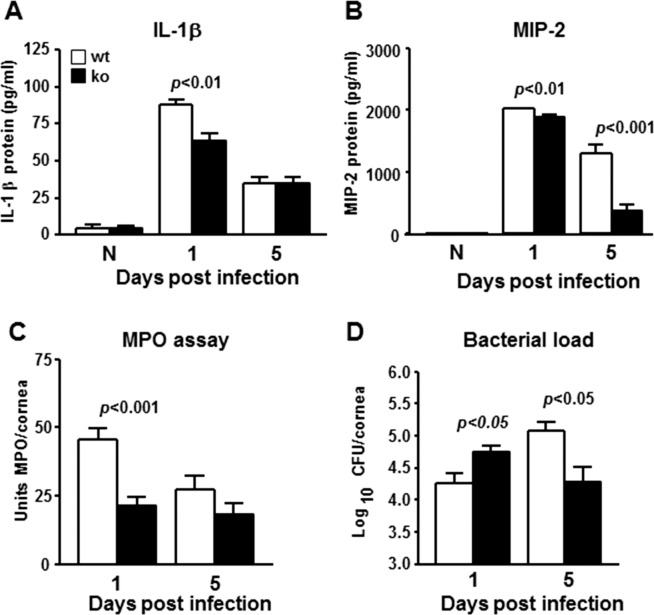
Methods: Eight-week-old miR-183/96/182 knockout (ko) mice and their wild-type littermates (wt) were used. The central cornea of anesthetized mice was scarred and infected with Pseudomonas aeruginosa (PA), strain 19660. Corneal disease was graded at 1, 3, and 5 days postinfection (dpi). Corneal RNA was harvested for quantitative RT-PCR. Polymorphonuclear neutrophils (PMN) were enumerated by myeloperoxidase assays; the number of viable bacteria was determined by plate counts, and ELISA assays were performed to determine cytokine protein levels. A macrophage (Mϕ) cell line and elicited peritoneal PMN were used for in vitro functional assays.
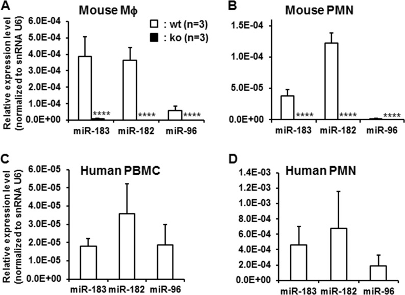
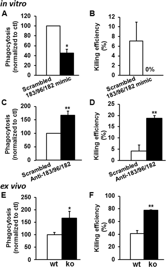
Results: MicroRNA-183/96/182 is expressed in the cornea, and in Mϕ and PMN of both mice and humans. Inactivation of miR-183/96/182 resulted in decreased corneal nerve density compared with wt mice. Overexpression of miR-183/96/182 in Mϕ decreased, whereas knockdown or inactivation of miR-183/96/182 in Mϕ and PMN increased their capacity for phagocytosis and intracellular killing of PA. In PA-infected corneas, ko mice showed decreased proinflammatory neuropeptides such as substance P and chemoattractant molecules, MIP-2, MCP1, and ICAM1; decreased number of PMN at 1 and 5 dpi; increased viable bacterial load at 1 dpi, but decreased at 5 dpi; and markedly decreased corneal disease.
Conclusions: MicroRNA-183/96/182 modulates the corneal response to bacterial infection through its regulation of corneal innervation and innate immunity.
Figures
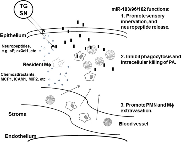
 , sensory nerve ending;
  , PMN;
  , Mϕ;
, Mϕ;  , PA;
, PA;  , neuropeptides and cytokines.
Similar articles
-
Prophylactic Knockdown of the miR-183/96/182 Cluster Ameliorates Pseudomonas aeruginosa-Induced Keratitis.Invest Ophthalmol Vis Sci. 2021 Dec 1;62(15):14. doi: 10.1167/iovs.62.15.14. Invest Ophthalmol Vis Sci. 2021. PMID: 34919120 Free PMC article.
-
Cathelicidin-deficient (Cnlp -/- ) mice show increased susceptibility to Pseudomonas aeruginosa keratitis.Invest Ophthalmol Vis Sci. 2007 Oct;48(10):4498-508. doi: 10.1167/iovs.07-0274. Invest Ophthalmol Vis Sci. 2007. PMID: 17898271 Free PMC article.
-
Interferon regulatory factor-1 in flagellin-induced reprogramming: potential protective role of CXCL10 in cornea innate defense against Pseudomonas aeruginosa infection.Invest Ophthalmol Vis Sci. 2013 Nov 15;54(12):7510-21. doi: 10.1167/iovs.13-12453. Invest Ophthalmol Vis Sci. 2013. PMID: 24130180 Free PMC article.
-
IL-33 shifts macrophage polarization, promoting resistance against Pseudomonas aeruginosa keratitis.Invest Ophthalmol Vis Sci. 2010 Mar;51(3):1524-32. doi: 10.1167/iovs.09-3983. Epub 2009 Nov 5. Invest Ophthalmol Vis Sci. 2010. PMID: 19892870 Free PMC article.
-
Corneal response to Pseudomonas aeruginosa infection.Prog Retin Eye Res. 2004 Jan;23(1):1-30. doi: 10.1016/j.preteyeres.2003.10.002. Prog Retin Eye Res. 2004. PMID: 14766315 Review.
Cited by
-
Challenges of corneal infections.Expert Rev Ophthalmol. 2016;11(4):285-297. doi: 10.1080/17469899.2016.1203254. Epub 2016 Jun 30. Expert Rev Ophthalmol. 2016. PMID: 28090214 Free PMC article.
-
Deciphering of Adult Glioma Vulnerabilities through Expression Pattern Analysis of GABA, Glutamate and Calcium Neurotransmitter Genes.J Pers Med. 2022 Apr 14;12(4):633. doi: 10.3390/jpm12040633. J Pers Med. 2022. PMID: 35455749 Free PMC article.
-
Role of microRNAs in Immune Regulation with Translational and Clinical Applications.Int J Mol Sci. 2024 Feb 5;25(3):1942. doi: 10.3390/ijms25031942. Int J Mol Sci. 2024. PMID: 38339220 Free PMC article. Review.
-
MicroRNAs in infectious diseases: potential diagnostic biomarkers and therapeutic targets.Clin Microbiol Rev. 2023 Dec 20;36(4):e0001523. doi: 10.1128/cmr.00015-23. Epub 2023 Nov 1. Clin Microbiol Rev. 2023. PMID: 37909789 Free PMC article. Review.
-
miRNAs reshape immunity and inflammatory responses in bacterial infection.Signal Transduct Target Ther. 2018 May 25;3:14. doi: 10.1038/s41392-018-0006-9. eCollection 2018. Signal Transduct Target Ther. 2018. PMID: 29844933 Free PMC article.
References
-
- Kerschensteiner M,, Meinl E,, Hohlfeld R. Neuro-immune crosstalk in CNS diseases. Neuroscience. 2009; 158: 1122–1132. - PubMed
-
- Steinman L. Elaborate interactions between the immune and nervous systems. Nat Immunol. 2004; 5: 575–581. - PubMed
-
- Brogden KA,, Guthmiller JM,, Salzet M,, Zasloff M. The nervous system and innate immunity: the neuropeptide connection. Nat Immunol. 2005; 6: 558–564. - PubMed
-
- Tsui H,, Razavi R,, Chan Y,, Yantha J,, Dosch HM. ‘Sensing' autoimmunity in type 1 diabetes. Trends Mol Med. 2007; 13: 405–413. - PubMed
-
- Razavi R,, Chan Y,, Afifiyan FN,, et al. TRPV1+ sensory neurons control beta cell stress and islet inflammation in autoimmune diabetes. Cell. 2006; 127: 1123–1135. - PubMed
Publication types
MeSH terms
Substances
Grants and funding
LinkOut - more resources
Full Text Sources
Other Literature Sources
Molecular Biology Databases
Research Materials
Miscellaneous
