. 2016 Apr 1;14(4):e1002427.
doi: 10.1371/journal.pbio.1002427. eCollection 2016 Apr.

Excessive Osteocytic Fgf23 Secretion Contributes to Pyrophosphate Accumulation and Mineralization Defect in Hyp Mice

Affiliations

Excessive Osteocytic Fgf23 Secretion Contributes to Pyrophosphate Accumulation and Mineralization Defect in Hyp Mice

Sathish K Murali et al. PLoS Biol. .

Abstract

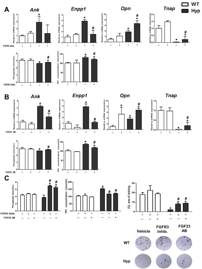
X-linked hypophosphatemia (XLH) is the most frequent form of inherited rickets in humans caused by mutations in the phosphate-regulating gene with homologies to endopeptidases on the X-chromosome (PHEX). Hyp mice, a murine homologue of XLH, are characterized by hypophosphatemia, inappropriately low serum vitamin D levels, increased serum fibroblast growth factor-23 (Fgf23), and osteomalacia. Although Fgf23 is known to be responsible for hypophosphatemia and reduced vitamin D hormone levels in Hyp mice, its putative role as an auto-/paracrine osteomalacia-causing factor has not been explored. We recently reported that Fgf23 is a suppressor of tissue nonspecific alkaline phosphatase (Tnap) transcription via FGF receptor-3 (FGFR3) signaling, leading to inhibition of mineralization through accumulation of the TNAP substrate pyrophosphate. Here, we report that the pyrophosphate concentration is increased in Hyp bones, and that Tnap expression is decreased in Hyp-derived osteocyte-like cells but not in Hyp-derived osteoblasts ex vivo and in vitro. In situ mRNA expression profiling in bone cryosections revealed a ~70-fold up-regulation of Fgfr3 mRNA in osteocytes versus osteoblasts of Hyp mice. In addition, we show that blocking of increased Fgf23-FGFR3 signaling with anti-Fgf23 antibodies or an FGFR3 inhibitor partially restored the suppression of Tnap expression, phosphate production, and mineralization, and decreased pyrophosphate concentration in Hyp-derived osteocyte-like cells in vitro. In vivo, bone-specific deletion of Fgf23 in Hyp mice rescued the suppressed TNAP activity in osteocytes of Hyp mice. Moreover, treatment of wild-type osteoblasts or mice with recombinant FGF23 suppressed Tnap mRNA expression and increased pyrophosphate concentrations in the culture medium and in bone, respectively. In conclusion, we found that the cell autonomous increase in Fgf23 secretion in Hyp osteocytes drives the accumulation of pyrophosphate through auto-/paracrine suppression of TNAP. Hence, we have identified a novel mechanism contributing to the mineralization defect in Hyp mice.


Conflict of interest statement

The authors have declared that no competing interests exist.

Figures

Fig 1
Fig 1. PPi concentration is increased in femurs of Hyp mice.
(A) Serum calcium, phosphate, ALP activity, and intact Fgf23 in 3-mo-old male WT and Hyp mice. (B) Von Kossa/McNeal staining of 3-μm-thick undecalcified plastic sections of distal femurs from 3-mo-old male WT and Hyp mice and quantification of mean area of osteocytic lacunae. (C) Quantification of OPN protein expression by western blotting of proteins isolated from femurs (upper panels) and immunohistochemical staining of OPN protein expression in femoral cortical bone (lower panels) in 3-mo-old male WT and Hyp mice. (D) PPi concentration in extracts of whole femurs from 3-mo-old male WT and Hyp mice. Each data point is the mean ± standard deviation (SD) of four mice. Individual values are given in S1 Data. *, p < 0.05 versus WT.
Fig 2
Fig 2. Osteocyte-rich cell fractions isolated from Hyp femurs show increased mRNA expression of PPi-regulating genes and decreased mRNA expression of Tnap ex vivo.
(A) mRNA abundance of the osteoblast-specific gene Ocn and of the osteocyte-specific gene Sost in total RNA isolated from cell fractions harvested by sequential digestion from femurs of 3-mo-old male WT and Hyp mice. (B–C) Ank, Enpp1, Opn, Fgf23, Tnap, Fgfr1, and Fgfr3 mRNA abundance in total RNA isolated from osteoblast-rich fractions F-3 to F-5 (B) and from osteocyte-rich fractions F-6/7 to F-8/9 (C) harvested by sequential digestion from femurs of 3-mo-old male WT and Hyp mice. (D) TNAP protein expression in pooled osteoblast- and osteocyte-rich fractions harvested by sequential digestion from femurs of 3-mo-old male WT and Hyp mice. Each data point is the mean ± SD of four samples from four different mice. Individual values are given in S1 Data. *, p < 0.05 versus F-3 in A; *, p < 0.05 versus WT in B–C; *, p < 0.05 versus WT F-3/4/5 in D.
Fig 3
Fig 3. Osteocyte-like cells isolated from Hyp mice display decreased Tnap mRNA expression together with PPi accumulation in vitro.
(A) mRNA abundance of the osteoblast-specific gene Ocn and of the osteocyte-specific gene Sost in calvarial cells isolated from newborn WT and Hyp mice and differentiated for 0–22 d (D0–D22). (B–C) mRNA abundance of Ank, Enpp1, Opn, Fgf23, Tnap, Fgfr1, and Fgfr3 as well as concentration of Pi, PPi, and intact Fgf23 in cell culture supernatant, percentage NBT/BCIP-stained area, and TNAP protein expression in calvarial cells isolated from newborn WT and Hyp mice and differentiated for 12 d (differentiated osteoblasts) (B) or 22 d (osteocyte-like cells) (C). Each data point is the mean ± SD of triplicates from four different animals. Individual values are given in S1 Data. *, p < 0.05 versus D0 in A; *, p < 0.05 versus WT in B and C.
Fig 4
Fig 4. Decreased TNAP enzyme activity and up-regulated expression of Fgfr3 mRNA in Hyp osteocytes in vivo.
(A) Histochemical TNAP staining in undecalcified sections of distal femurs from WT and Hyp mice and quantification of relative fluorescence. Top panels for each genotype show endocortical bone surface, bottom panels cortical bone. Each data point is the mean ± SD of at least four mice. (B) In situ mRNA expression profiling of osteoblasts and osteocytes harvested by laser capture microdissection (LCM) in 4-μm-thick distal femoral cryosections. Each data point is the mean ± SD of three mice. Individual values are given in S1 Data. *, p < 0.05 versus WT osteocytes in A; *, p < 0.05 versus WT osteoblasts, #, p < 0.05 versus WT osteocytes in B.
Fig 5
Fig 5. Inhibition of Fgf23-FGFR3 signaling increases TNAP expression and lowers PPi concentration in Hyp-derived osteocyte-like cells.
(A–B) Ank, Enpp1, Opn and Tnap mRNA abundance as well as Pi and PPi concentration in cell culture supernatant of calvarial osteocyte-like cells differentiated for 22 d and treated for 24 h with a FGFR3 inhibitor (A) or anti-FGF23 antibody (FGF23 AB) (B). (C) Phosphate and PPi concentration in cell culture supernatant as well as percent NBT/BCIP-stained area in cultures of calvarial osteocyte-like cells differentiated for 22 d, and treated daily over 4 d with a FGFR3 inhibitor or FGF23 AB. Each data point is the mean ± SD of four experimental samples. Individual values are given in S1 Data. *, p < 0.05 versus vehicle-treated WT cells, #, p < 0.05 versus vehicle-treated Hyp cells.
Fig 6
Fig 6. rFGF23 administration suppresses Tnap mRNA expression and increases PPi concentration in WT osteocyte-like cells and bones of WT mice.
(A) mRNA abundance of Ank, Enpp1, and Tnap as well as PPi concentration in cell culture supernatant in WT calvarial osteocyte-like cells differentiated for 22 d, and subsequently treated with rFGF23 for 24 h. (B) mRNA abundance of Ank, Enpp1, and Tnap in total RNA isolated from whole femurs and PPi concentration in extracts of whole tibiae from 3-mo-old male WT mice treated with rFGF23 or vehicle for 5 d. Each data point is the mean ± SD of at least four experimental samples in A, and of at least five mice in B. Individual values are given in S1 Data. *, p < 0.05 versus vehicle.
Fig 7
Fig 7. Bone specific deletion of Fgf23 rescues suppressed TNAP activity in Hyp mice.
Histochemical TNAP staining in bone sections from WT, Hyp, and Hyp/Fgf23 Δ/flox /Col2.3 cre+ mice and quantification of relative fluorescence in osteoblasts and osteocytes. Top panels for each genotype represent endocortical bone surface, bottom panels cortical bone. Each data point is the mean ± SD of at least four mice. Individual values are given in S1 Data. *, p < 0.05 versus WT; #, p < 0.05 versus Hyp.
Fig 8
Fig 8. Proposed model of PPi accumulation through increased Fgf23-FGFR3 signaling in Hyp osteocytes.
Fgf23 secreted into the extracellular fluid binds to FGFR3 and acts as a transcriptional suppressor of Tnap. TNAP is a central molecule in the mineralization process and favors mineralization through hydrolyzing PPi, thus providing Pi for mineralization. Fgf23 secretion is up-regulated in osteoblasts and especially osteocytes in Hyp bones, relative to WT osteoblasts and osteocytes. FGFR3 expression is reduced in Hyp compared with WT osteoblasts. However, differentiation of osteoblasts into osteocytes is associated with a distinct up-regulation of FGFR3 expression especially in Hyp bones. Together with the increased secretion of Fgf23, the upregulation in FGFR3 leads to autocrine/paracrine suppression of TNAP activity in Hyp osteocytes but not osteoblasts, causing accumulation of PPi and inhibition of bone mineralization in osteocyte lacunae.

References

    1. Francis F, Hennig S, Korn B, Reinhardt R, de Jong P, Poustka A, et al. A gene (PEX) with homologies to endopeptidases is mutated in patients with X-linked hypophosphatemic rickets. Nat Genet. 1995;11: 130–6. - PubMed
    1. Holm IA, Huang X, Kunkel LM. Mutational analysis of the PEX gene in patients with X-linked hypophosphatemic rickets. Am J Hum Genet. 1997;60: 790–7. - PMC - PubMed
    1. Rowe PS, Oudet CL, Francis F, Sinding C, Pannetier S, Econs MJ, et al. Distribution of mutations in the PEX gene in families with X-linked hypophosphataemic rickets (HYP). Hum Mol Genet. 1997;6: 539–49. - PubMed
    1. Beck L, Soumounou Y, Martel J, Krishnamurthy G, Gauthier C, Goodyer CG, et al. Pex/PEX tissue distribution and evidence for a deletion in the 3' region of the Pex gene in X-linked hypophosphatemic mice. J Clin Invest. 1997;99: 1200–9. - PMC - PubMed
    1. Du L, Desbarats M, Viel J, Glorieux FH, Cawthorn C, Ecarot B. cDNA cloning of the murine Pex gene implicated in X-linked hypophosphatemia and evidence for expression in bone. Genomics. 1996;36: 22–8. - PubMed

Publication types

MeSH terms

LinkOut - more resources