. 2016 Jun;157(6):2432-46.
doi: 10.1210/en.2015-1942. Epub 2016 Apr 1.

Endometrial Stromal Decidualization Responds Reversibly to Hormone Stimulation and Withdrawal

Affiliations

Endometrial Stromal Decidualization Responds Reversibly to Hormone Stimulation and Withdrawal

Jie Yu et al. Endocrinology. 2016 Jun.

Abstract

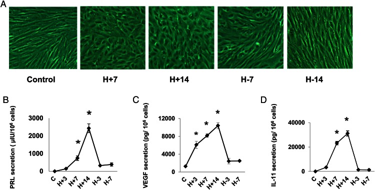
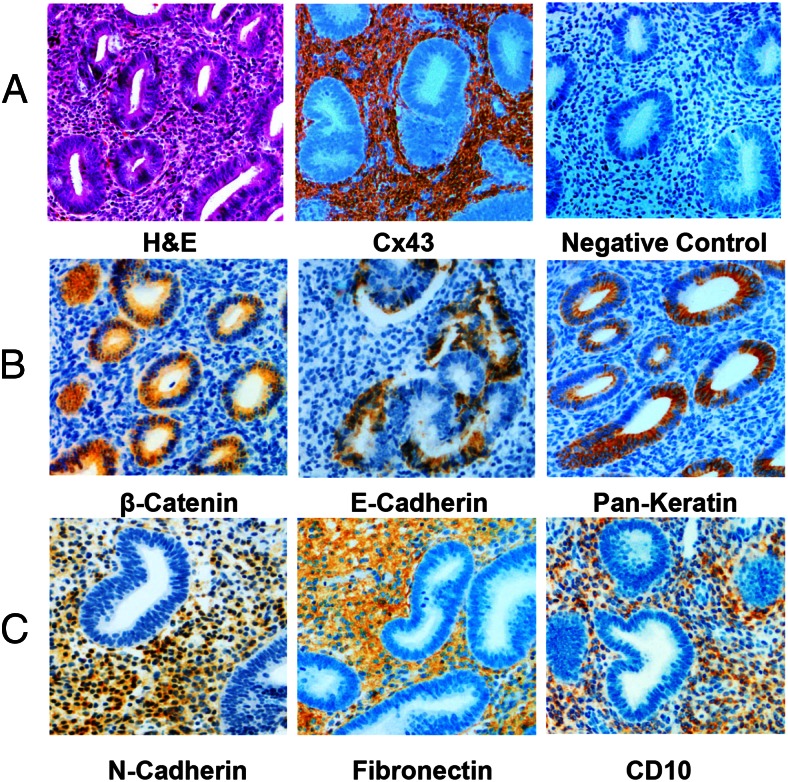
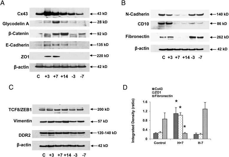
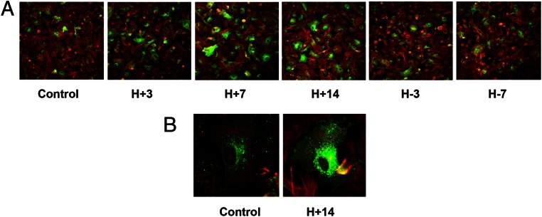
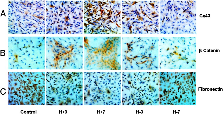
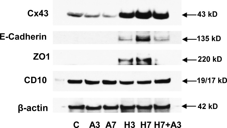
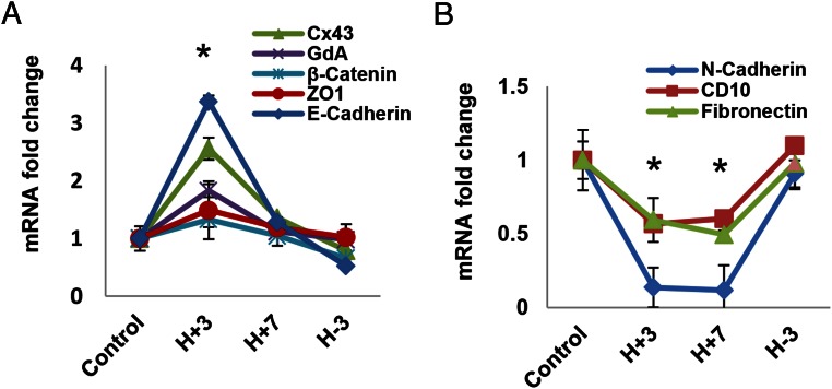
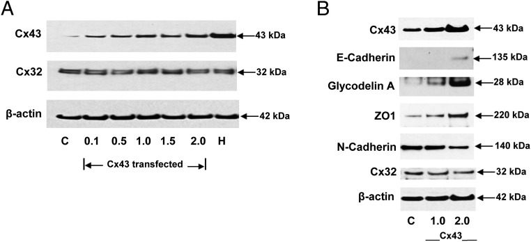
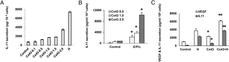
Human endometrial stromal decidualization is required for embryo receptivity, angiogenesis, and placentation. Previous studies from our laboratories established that connexin (Cx)-43 critically regulates endometrial stromal cell (ESC) differentiation, whereas gap junction blockade prevents it. The current study evaluated the plasticity of ESC morphology and Cx43 expression, as well as other biochemical markers of cell differentiation, in response to decidualizing hormones. Primary human ESC cultures were exposed to 10 nM estradiol, 100 nM progesterone, and 0.5 mM cAMP for up to 14 days, followed by hormone withdrawal for 14 days, mimicking a biphasic ovulatory cycle. Reversible differentiation was documented by characteristic changes in cell shape. Cx43 was reversibly up- and down-regulated after the estradiol, progesterone, and cAMP treatment and withdrawal, respectively, paralleled by fluctuations in prolactin, vascular endothelial growth factor, IL-11, and glycodelin secretion. Markers of mesenchymal-epithelial transition (MET), and its counterpart epithelial-mesenchymal transition, followed reciprocal patterns corresponding to the morphological changes. Incubation in the presence of 18α-glycyrrhetinic acid, an inhibitor of gap junctions, partially reversed the expression of decidualization and MET markers. In the absence of hormones, Cx43 overexpression promoted increases in vascular endothelial growth factor and IL-11 secretion, up-regulated MET markers, and reduced N-cadherin, an epithelial-mesenchymal transition marker. The combined results support the hypothesis that Cx43-containing gap junctions and endocrine factors cooperate to regulate selected biomarkers of stromal decidualization and MET and suggest roles for both phenomena in endometrial preparation for embryonic receptivity.


Figures

Figure 1.
Figure 1.
A, Hormone-induced changes in ESC morphology. ESCs were cultured without added hormones (control) or with 10 nM E2, 100 nM P4, and 0.5 mM dibutyryl cAMP (E/P/c) for 7 days (H+7). ESCs incubated with E/P/c for 14 days showed maximal decidualization morphology (H+14). When ESCs were incubated for 14 days with E/P/c and then withdrawn from the hormones for 7 days, the cells became more fibroblastic in appearance (H-7). After 7 further days of hormone withdrawal, the cell shape approached that of controls (H-14). B–D, Hormone-induced changes in biomarker secretion (ELISA). ESCs were cultured for up to 14 days (H+14) with E/P/c as in panel A and then withdrawn from the decidualizing stimulus for up to 7 days (H-7). The secretion of prolactin (panel B), VEGF (panel C), and IL-11 (panel D), all determined by ELISA, had similar patterns. *, P < .05, ANOVA with Scheffé's post hoc tests (different from controls).
Figure 2.
Figure 2.
A–C, Histology and IHC of proliferative-phase endometrium. Biopsies of proliferative human endometrium were collected as described and stained with hematoxylin and eosin (H&E), Cx43 antibodies or nonimmune rabbit serum as a negative control (panel A). Epithelial proteins were identified by antibodies against β-catenin, E-cadherin, and pan-keratin (panel B). Stromal proteins were localized using antibodies against N-cadherin, fibronectin, and CD10 (panel C).
Figure 3.
Figure 3.
A, Hormone-induced changes in epithelial protein expression (Western blots). ESCs were cultured for up to 14 days (+14) with E/P/c and then withdrawn from the decidualizing stimulus for up to 7 days (−7) as in Figure 1. The individual kinetics of several different proteins were assessed by Western blotting with specific antibodies to each. The molecular mass of each band is denoted at the right and the common protein names or abbreviations are indicated at the left. Expression of β-actin was constant across the time course. B, Hormone-induced changes in stromal protein expression (Western blots). ESCs were cultured for up to 14 days (+14) with E/P/c and hormone withdrawn for up to 7 days (−7) as in Figure 1. The molecular mass of each band is denoted at the right and the common protein names or abbreviations are indicated at the left. C, Some EMT biomarkers remain stable despite decidual hormone stimulation and withdrawal. ESCs were cultured for up to 14 days (+14) with E/P/c and hormone withdrawn for up to 7 days (−7) as in Figure 1. The molecular mass of each band is denoted at the right and the common protein names or abbreviations of EMT markers are indicated at the left. D, Quantitative changes in Cx43, ZO1, and fibronectin. Densitometry of Western blot bands from at least three biological replicates at each time point (control, no hormones; H+7, E/P/c stimulation for 7 d; H-7, E/P/c stimulation for 7 d followed by 7 d hormone withdrawal) was performed. The densitometry of each protein was normalized to the intensity of corresponding β-actin bands was calculated as an integrated density (ratio) and subjected to statistical analysis. *, P < .05, ANOVA with Scheffé's post hoc tests (different from control). C, control.
Figure 4.
Figure 4.
A and B, Immunofluorescence ICC confirms hormone-induced changes in Cx43 protein expression. Immunofluorescence ICC in ESC monolayers showed Cx43 up- and down-regulation after the E/P/c-induced decidualization (H+3, H+7, H+14) and withdrawal (H-3, H-7, panel A). Green fluorescence represents Cx43 and red fluorescence is β-actin. Panel B shows a high-magnification view (×480) demonstrating intracellular distribution of Cx43 (green) under control conditions and after 14 days of hormone stimulation.
Figure 5.
Figure 5.
Immunoperoxidase ICC confirms hormone-induced changes in Cx43, MET, and EMT protein expression. Immunoperoxidase ICC in ESC monolayers showed Cx43 up- and down-regulation after E/P/c-induced decidualization (H+3, H+7) and withdrawal (H-3, H-7, panel A). Sequential up- and down-regulation of the epithelial marker β-catenin was associated with hormone exposure and withdrawal (panel B). Conversely, the stromal marker fibronectin showed concomitant down- and up-regulation, respectively (panel C). Magnification, ×200.
Figure 6.
Figure 6.
Gap junction inhibition reverses decidualized phenotype and MET changes. Incubation in the presence of a GJIC inhibitor (50 μM 8α-AGA [A]) for 3 or 7 days resulted in a decrease in Cx43 under basal (A3, A7) conditions. E/P/c treatment for 3 or 7 days (H3, H7) increased Cx43, but the latter effect was reversed when AGA also was present from day 4 to day 7 (H7+A3). Similarly, the up-regulation of E-cadherin and ZO1 after 3 and 7 days of E/P/c was blocked when AGA also was present from day 4 to day 7 (H7+A3). CD10 was reduced subtly after 3 or 7 days of E/P/c, with some reversal when AGA also was present from day 4 to day 7 (H7+A3). Expression of β-actin was stable across time and treatments. C, control.
Figure 7.
Figure 7.
Hormone-induced changes in mRNA expression (qRT-PCR). ESCs were cultured for 3 or 7 days (H+3, H+7) with E/P/c and then withdrawn from the decidualizing stimulus for up to 3 days (H-3), similar to the conditions above but for shorter intervals, to examine kinetics of accumulation of mRNAs corresponding to the proteins in Figure 3. A, Cx43, GdA, β-catenin, and E-cadherin were all different from control at H+3 days. ZO1 showed a similar trend but did not meet significance. B, N-cadherin, CD10, and fibronectin all differed from control at H+3 and H+7 days. *, P < .05, ANOVA with Scheffé's post hoc test (different from control).
Figure 8.
Figure 8.
A, Transfection of Cx43 expression vectors dose responsively induces specific gap junction proteins. To optimize Cx43 transfection, dose-response experiments with increasing vector concentrations (0.1–2.0 μg per 6 cm dish) were performed. The EC50 was determined to be approximately 1.0 μg per 6-cm dish, which yielded less Cx43 protein accumulation than that achieved after 7 days of E/P/c treatment (H). Note that neither Cx43 overexpression nor E/P/c treatment had any effect on Cx32 or β-actin expression. B, Transfection of Cx43 expression vectors dose responsively increases MET markers and decreases N-cadherin in the absence of hormone treatment. Transfection of 1.0 and 2.0 μg per 6 cm dish Cx43 vectors resulted in increases in Cx43. Western blots of MET markers (E-cadherin, GdA, and ZO1) also showed increases. By contrast, the stromal marker N-cadherin was reduced when Cx43 was overexpressed. All experiments were performed in the absence of hormones. No effects on Cx32 or β-actin were observed. C, control.
Figure 9.
Figure 9.
A and B, Overexpression of Cx43 induces ESC up-regulation of IL-11 in the absence of hormone treatment. Transgenic expression of Cx43 dose responsively up-regulates IL-11 secretion, with 2.0 μg per 6-cm dish approaching approximately 50% the magnitude of secretion after 7 days of exposure to E/P/c (panel A). The combination of recombinant Cx43 and decidual hormones have a more than additive effect on IL-11 secretion (panel B). *, P < .05, t tests (different from corresponding controls). C, Overexpression of Cx43 up-regulates VEGF and IL-11 production in ESCs. Transfection of 1.0 μg Cx43 vector DNA per 6-cm dish increased VEGF secretion by 2.3-fold and IL-11 by 7.2-fold, but these responses were lower than those induced by E/P/c for 7 days (H), *, P < .05, t tests (different from control). Additive effects were noted when transfection was combined with E/P/c treatment (Cx43+H), **, P < .05, t tests (different from corresponding Cx43 transfection controls [Cx43]).

References

    1. Martin RD. The evolution of human reproduction: a primatological perspective. Am J Phys Anthropol Suppl. 2007;45:59–84. - PubMed
    1. Gargett CE, Masuda H. Adult stem cells in the endometrium. Mol Hum Reprod. 2010;16:818–834. - PubMed
    1. Brosens JJ, Wilson MS, Lam EW. FOXO transcription factors: from cell fate decisions to regulation of human female reproduction. Adv Exp Med Biol. 2009;665:227–241. - PubMed
    1. Ramathal CY, Bagchi IC, Taylor RN, Bagchi MK. Endometrial decidualization: of mice and men. Semin Reprod Med. 2010;28:17–26. - PMC - PubMed
    1. Gellersen B, Brosens IA, Brosens JJ. Decidualization of the human endometrium: mechanisms, functions, and clinical perspectives. Semin Reprod Med. 2007;25:445–453. - PubMed

Publication types