. 2016 Apr 1;12(4):e1005521.
doi: 10.1371/journal.ppat.1005521. eCollection 2016 Apr.

Endoplasmic Reticulum Stress Induced Synthesis of a Novel Viral Factor Mediates Efficient Replication of Genotype-1 Hepatitis E Virus

Affiliations

Endoplasmic Reticulum Stress Induced Synthesis of a Novel Viral Factor Mediates Efficient Replication of Genotype-1 Hepatitis E Virus

Vidya P Nair et al. PLoS Pathog. .

Abstract

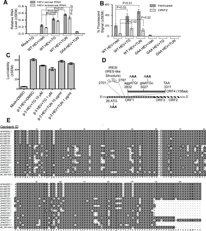
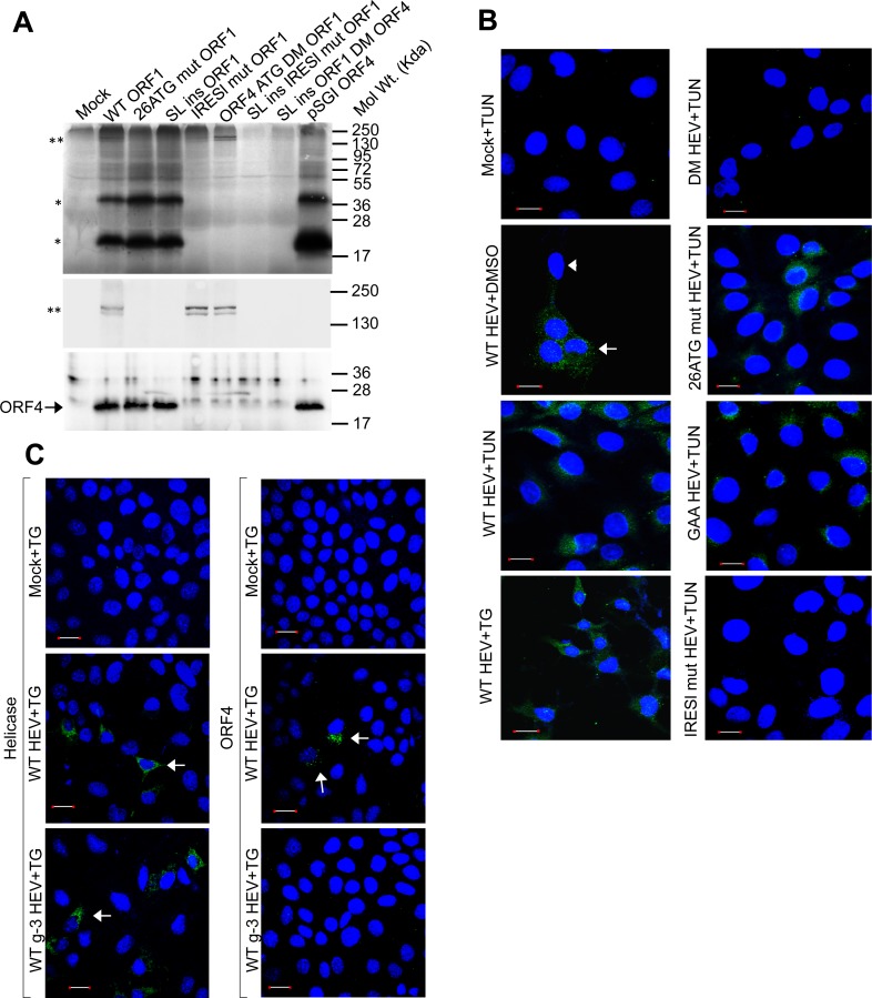
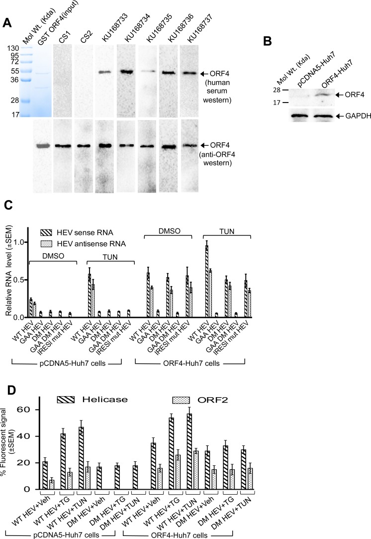
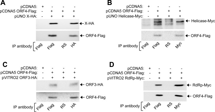
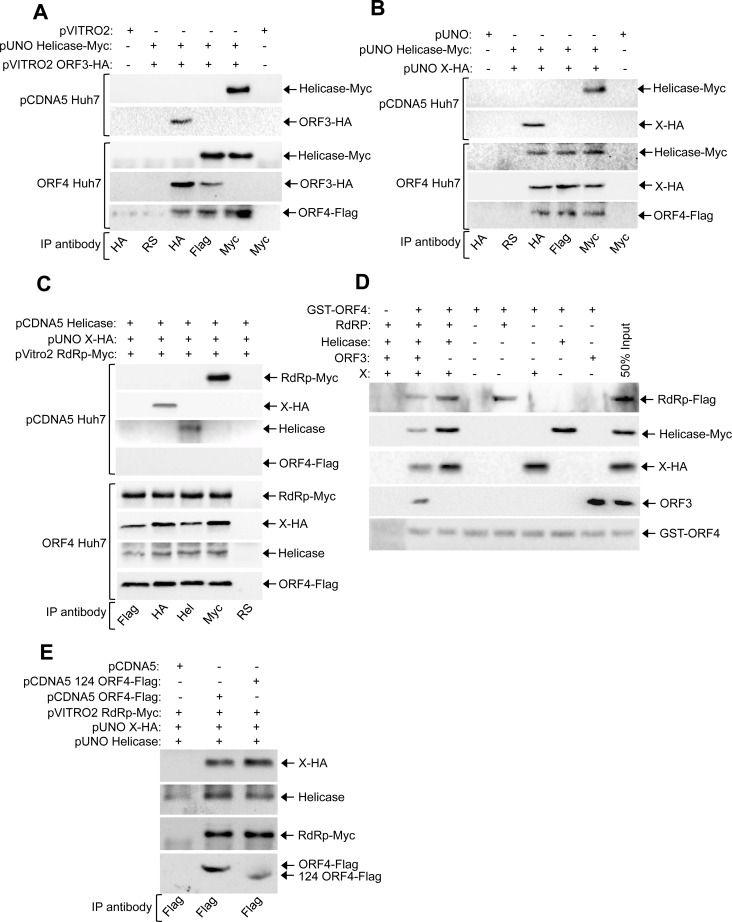
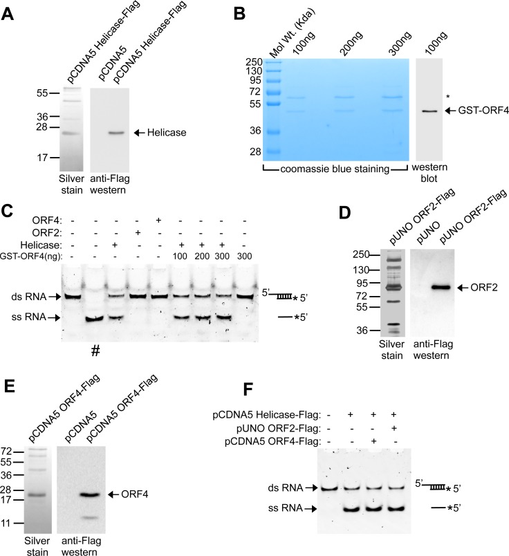
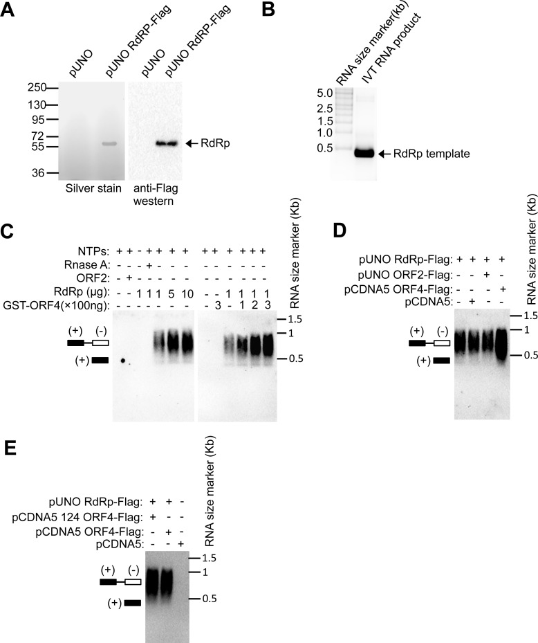
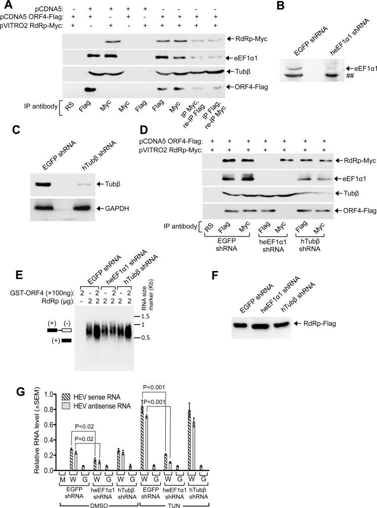
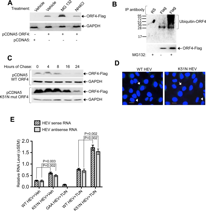
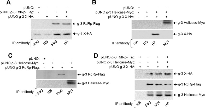
Hepatitis E virus (HEV) causes acute hepatitis in many parts of the world including Asia, Africa and Latin America. Though self-limiting in normal individuals, it results in ~30% mortality in infected pregnant women. It has also been reported to cause acute and chronic hepatitis in organ transplant patients. Of the seven viral genotypes, genotype-1 virus infects humans and is a major public health concern in South Asian countries. Sporadic cases of genotype-3 and 4 infection in human and animals such as pigs, deer, mongeese have been reported primarily from industrialized countries. Genotype-5, 6 and 7 viruses are known to infect animals such as wild boar and camel, respectively. Genotype-3 and 4 viruses have been successfully propagated in the laboratory in mammalian cell culture. However, genotype-1 virus replicates poorly in mammalian cell culture and no other efficient model exists to study its life cycle. Here, we report that endoplasmic reticulum (ER) stress promotes genotype-1 HEV replication by inducing cap-independent, internal initiation mediated translation of a novel viral protein (named ORF4). Importantly, ORF4 expression and stimulatory effect of ER stress inducers on viral replication is specific to genotype-1. ORF4 protein sequence is mostly conserved among genotype-1 HEV isolates and ORF4 specific antibodies were detected in genotype-1 HEV patient serum. ORF4 interacted with multiple viral and host proteins and assembled a protein complex consisting of viral helicase, RNA dependent RNA polymerase (RdRp), X, host eEF1α1 (eukaryotic elongation factor 1 isoform-1) and tubulinβ. In association with eEF1α1, ORF4 stimulated viral RdRp activity. Furthermore, human hepatoma cells that stably express ORF4 or engineered proteasome resistant ORF4 mutant genome permitted enhanced viral replication. These findings reveal a positive role of ER stress in promoting genotype-1 HEV replication and pave the way towards development of an efficient model of the virus.


Conflict of interest statement

The authors have declared that no competing interests exists.

Figures

Fig 1
Fig 1. Tunicamycin and thapsigargin promote g-1 HEV replication.
(A) QRT-PCR of wild type (WT HEV) and replication deficient (GAA HEV) HEV in Huh7 cells transfected with in vitro synthesized genome. TG: thapsigargin, TUN: tunicamycin. Values are mean±SEM. (B) Quantitation of viral ORF1 (Helicase) and ORF2 expression in Huh7 cells, transfected with wild type (WT HEV) or replication deficient (GAA HEV) HEV genomic RNA and treated with the indicated compounds. Ten random fields of approximately 30 cells in each field were counted for helicase, ORF2, DAPI (nuclear stain) fluorescence and percentage ±SEM calculated. (C) Measurement of secreted Gaussia luciferase activity in the culture medium of Huh7 cells expressing in vitro transcribed HEV genotype-3 replicon RNA and treated as indicated. Values are mean±SEM. (D) HEV genome organisation. Numbers indicate nucleotide position from 5’-end. Mutated bases are in bold. (E) ClustalW alignment of ORF4 protein sequence of indicated g-1 HEV isolates.
Fig 2
Fig 2. Tunicamycin and thapsigargin induce ORF4 expression.
(A) Top: Autoradiogram showing TNT of indicated plasmids. Mock: empty vector, “**”: ORF1 protein, “*”: unknown proteins. Middle: samples from top resolved by 7% SDS-PAGE, followed by autoradiography. Bottom: samples from top resolved by 15% SDS-PAGE and western using anti-ORF4. (B) Immunofluorescence of ORF4 in Huh-7 cells transfected with indicated in vitro synthesized RNA. Scale: 20μm. Shown are merged images of nuclei (blue) and ORF4 (green). “→”: positive staining, “►”: unstained. (C) Immunofluorescence of Helicase and ORF4 in Huh-7 cells transfected with in vitro synthesized wild type HEV g-1 (WT HEV) or g-3 (WT g-3 HEV) genomic RNA. Scale: 20μm. Shown are merged images of nuclei (blue) and Helicase or ORF4 (green). “→”: positive staining.
Fig 3
Fig 3. ORF4 antibody is detected in HEV patients and its over expression enhances viral replication.
(A) Top: Purified GST-ORF4 stained with coomassie blue (left most) and western of equal aliquots of the same using healthy (CS1, CS2) and acute HEV infected (KU168733-KU168737) patient sera. Bottom: Top blots reprobed with anti-ORF4 antibody. (B) Western of whole cell extract from indicated cells using ORF4 and GAPDH antibodies. (C) QRT-PCR of sense and anti-sense RNA in pCDNA5-Huh7 and ORF4-Huh7 cells transfected with in vitro synthesized wild type (WT) or mutant HEV genome and treated as indicated.(D) Quantitation of viral ORF1 (helicase) and ORF2 expression in pCDNA5-Huh7 and ORF4-Huh7 cells, transfected with wildtype (WT HEV) or ORF4 expression deficient mutant (DM HEV) HEV genomic RNA and treated with the indicated compounds. Ten random fields of approximately 30 cells in each field were counted for helicase, ORF2, DAPI (nuclear stain) fluorescence and percentage ±SEM calculated.
Fig 4
Fig 4. An IRESl (Internal ribosome entry site-like) element located upstream of ORF4 coding sequence drives its translation independent of ORF1.
(A) Predicted secondary structure of IRESl using “mfold”. Cyan letters indicate core IRESl sequence. A, B, C: stem loops, A*: bulge. Substitutions that impair IRESl are in bold. (B) Organization of Dual luciferase reporter plasmid. (C) Dual luciferase reporter assay. Values are mean±SEM.
Fig 5
Fig 5. ORF4 interacts with multiple viral proteins.
(A) CoIP of ORF4 and X expressing Huh7 extract, immunoprecipitated and revealed using indicated antibodies. RS: Rabbit preimmune serum. (B) CoIP of ORF4 and Helicase expressing Huh7 extract, immunoprecipitated and revealed using indicated antibodies. (C) CoIP of ORF4 and ORF3 expressing Huh7 extract, immunoprecipitated and revealed using indicated antibodies. (D) CoIP of ORF4 and RdRp expressing Huh7 extract, immunoprecipitated and revealed using indicated antibodies.
Fig 6
Fig 6. ORF4 mediates the assembly of a protein complex consisting of viral RdRp, Helicase and X.
(A) CoIP and western of pCDNA5-Huh7 and ORF4-Huh7 stable cell line extract transiently expressing myc-tagged helicase and HA-tagged ORF3 proteins, immunoprecipitated and revealed using indicated antibodies. RS: rabbit pre immune serum. (B) CoIP and western of pCDNA5-Huh7 and ORF4-Huh7 stable cell line extract transiently expressing myc-tagged helicase and HA-tagged X proteins, immunoprecipitated and revealed using indicated antibodies. (C) CoIP and western of pCDNA5-Huh7 and ORF4-Huh7 stable cell line extract transiently expressing helicase, X and RdRp, immunoprecipitated and revealed using indicated antibodies. (D) Pull down of ORF4 interacting proteins using GST-ORF4 as bait, revealed using indicated antibodies. Lane 1 contains GST as bait. (E) Pull down of full length ORF4 (pCDNA5 ORF4-Flag) and 1-124aa mutant ORF4 (pCDNA5 124 ORF4-Flag) interacting proteins using ORF4-Flag as bait, revealed using indicated antibodies.
Fig 7
Fig 7. ORF4 does not affect RNA strand displacement ability of g-1 HEV Helicase.
(A) Left panel: SDS-PAGE and Silver staining of Flag antibody immunoprecipitated samples from Huh7 cells transiently transfected with pCDNA5 Helicase-flag vector. Right panel: Western of pCDNA5 and pCDNA5 Helicase-flag vectors using anti-Flag antibody. (B) Left panel: SDS PAGE and Coomassie blue staining of indicated amount of purified bacterial GST-ORF4 protein. “*” indicates copurified unrelated protein band. Right panel: Western of 100ng purified GST-ORF4 protein using anti-ORF4 antibody. (C) Assay of RNA strand displacement by Helicase in the presence of GST-ORF4. “#”: heat denatured dsRNA. (D) Left panel: SDS-PAGE and Silver staining of Flag antibody immunoprecipitated samples from Huh7 cells transiently transfected with pUNO ORF2-flag vector. Right panel: Western of pUNO and pUNO ORF2-flag vector using anti-Flag antibody. (E) Left panel: SDS-PAGE and Silver staining of Flag antibody immunoprecipitated samples from Huh7 cells transiently transfected with pCDNA5 ORF4-flag vector. Right panel: Western of pCDNA5 and pCDNA5 ORF4-flag vector using anti-Flag antibody. (F) RNA strand displacement assay demonstrating the effect of Huh7 cell expressed ORF4 on HEV helicase enzymatic activity. Schematic at the right represents the structure of dsRNA and ssRNA.
Fig 8
Fig 8. ORF4 promotes viral RdRp activity.
(A) Left panel: SDS-PAGE and Silver staining of Flag antibody immunoprecipitated samples from Huh7 cells transiently transfected with pUNO empty vector or pUNO RdRp-flag vector. Right panel: Western of left panel samples using anti-Flag antibody. (B) Formaldehyde Agarose gel electrophoresis followed by staining with Ethidium bromide to detect in vitro transcribed RNA (IVT RNA product) used as template in RdRp assay. (C) RdRp assay using purified RdRp and GST-ORF4. Schematic illustrates position of dsRNA (+,-) and ss RNA. NTPs: Nucleotide triphosphate mix. (D) RdRp assay using purified RdRp, ORF2 and ORF4 proteins. (E) RdRp assay using purified RdRp, ORF2 and 124 ORF4 (1-124aa) proteins.
Fig 9
Fig 9. eEF1α1 bridges the interaction between ORF4 and RdRp.
(A) CoIP of RdRp, ORF4, eEF1α1 and Tubulin β (TUBβ) in Huh7 cells. (B) Western of Huh7 cells expressing EGFP or heEF1α1 shRNA. “##”: nonspecific band. (C) Western of Huh7 cells expressing EGFP or hTubβ shRNA (Top). Bottom: same blot reprobed with anti-GAPDH. (D) CoIP of Huh7 cells expressing EGFP, heEF1α1 and hTubβ shRNA. (E) HEV RdRp assay using EGFP, heEF1α1 and hTubβ shRNA expressing cells. (F) Anti-Flag western of Flag-affinity purified g-1 RdRp protein from Huh7 cells expressing g-1 RdRp-Flag and shRNAs against EGFP, eEF1α1 and Tubβ. (G) QRT-PCR of sense and antisense RNA of WT HEV (W) and GAA HEV (G) in Huh7 cells expressing EGFP, heEF1α1, hTubβ shRNA. M: Mock.
Fig 10
Fig 10. ORF4 is a target of host ubiquitin-proteasome machinery.
(A) Western blot using anti-Flag (top) and anti-GAPDH (bottom). (B) Western blot using anti-ubiquitin (top) and anti-Flag (bottom). (C) Anti-Flag western blot of Huh7 cells expressing WT and K51N mut ORF4, treated with cycloheximide (panel 1, 3). Same blots were reprobed with anti-GAPDH (panel 2, 4). (D) Immunofluorescence of ORF4 in Huh7 cells transfected with in vitro synthesized WT HEV or K51N HEV. Scale: 20μm. Shown are merged images of nuclei (blue) and ORF4 (green). “→”: positive staining, “►”: unstained. (E) QRT-PCR of HEV sense and anti-sense RNA from Huh7 cells transfected and treated as indicated. Veh: Vehicle, TUN:tunicamycin.
Fig 11
Fig 11. G-3 RdRp, X and Helicase associate with each other in Huh7 cells.
(A) CoIP and western of Huh7 cell extract transiently expressing g-3 RdRp and X, immunoprecipitated and revealed using indicated antibodies. (B) CoIP and western of Huh7 cell extract transiently expressing g-3 Helicase and X, immunoprecipitated and revealed using indicated antibodies. (C) CoIP and western of Huh7 cell extract transiently expressing g-3 RdRp and Helicase, immunoprecipitated and revealed using indicated antibodies. (D) CoIP and western of Huh7 cell extract transiently expressing g-3 RdRp, Helicase and X, immunoprecipitated and revealed using indicated antibodies.

References

    1. Ahmad I, Holla PR, Jameel S. Molecular virology of hepatitis E virus. Virus Res 2011;161(1): 47–58. 10.1016/j.virusres.2011.02.011 - DOI - PMC - PubMed
    1. Kamar N, Selves J, Mansuy MJ, Ouezzani L, Peron MJ, Guitard J et al. Hepatitis E Virus and chronic hepatitis in organ transplant recipients. N Engl J Med 2008;358(8): 811–17. 10.1056/NEJMoa0706992 - DOI - PubMed
    1. Khuroo MS, Teli MR, Skidmore S, Sofi MA, Khuroo MI. Incidence and severity of viral hepatitis in pregnancy. Am J Med 1981;70(2): 252–55. - PubMed
    1. Surjit M, Oberoi R, Kumar R, Lal SK. Enhanced α1 microglobulin secretion from hepatitis E virus ORF3 expressing human hepatoma cells is mediated by the Tumor susceptibility gene 101. J Biol Chem 2006;281(12): 8135–42. - PubMed
    1. Yamada K, Takahashi M, Hoshino Y, Takahashi H, Ichiyama K et al. ORF3 protein of hepatitis E virus is essential for virion release from infected cells. J Gen Virol 2009;90(Pt 8): 1880–91. 10.1099/vir.0.010561-0 - DOI - PubMed

Publication types