. 2016 Apr 12;113(15):4057-62.
doi: 10.1073/pnas.1524385113. Epub 2016 Mar 30.

A bacterial Argonaute with noncanonical guide RNA specificity

Affiliations

A bacterial Argonaute with noncanonical guide RNA specificity

Emine Kaya et al. Proc Natl Acad Sci U S A. .

Abstract

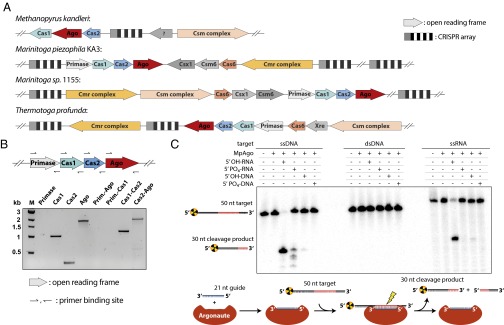
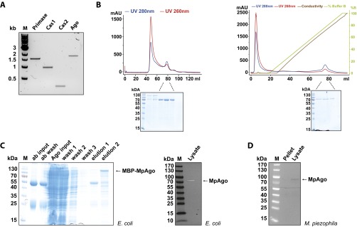
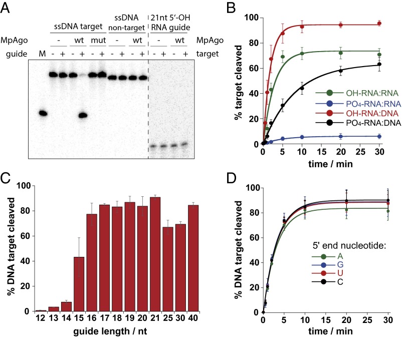
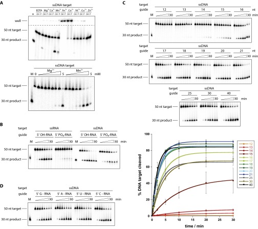
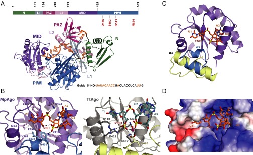
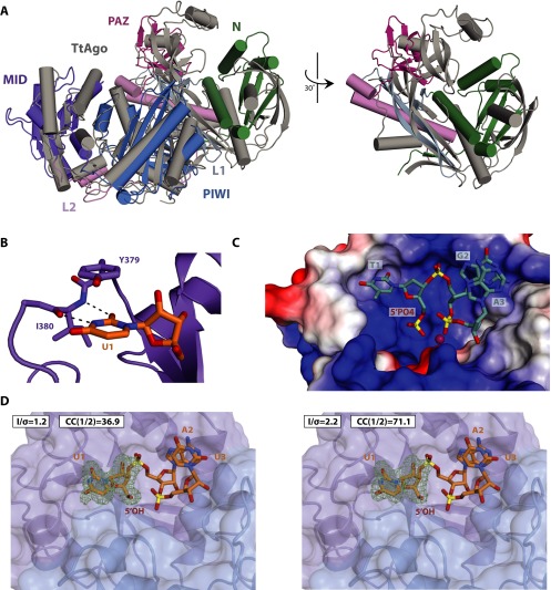
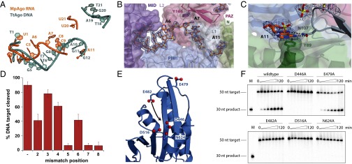
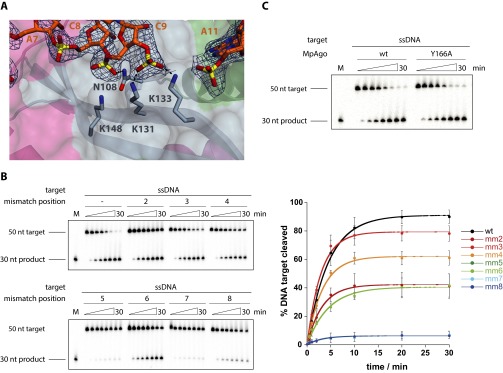
Eukaryotic Argonaute proteins induce gene silencing by small RNA-guided recognition and cleavage of mRNA targets. Although structural similarities between human and prokaryotic Argonautes are consistent with shared mechanistic properties, sequence and structure-based alignments suggested that Argonautes encoded within CRISPR-cas [clustered regularly interspaced short palindromic repeats (CRISPR)-associated] bacterial immunity operons have divergent activities. We show here that the CRISPR-associated Marinitoga piezophila Argonaute (MpAgo) protein cleaves single-stranded target sequences using 5'-hydroxylated guide RNAs rather than the 5'-phosphorylated guides used by all known Argonautes. The 2.0-Å resolution crystal structure of an MpAgo-RNA complex reveals a guide strand binding site comprising residues that block 5' phosphate interactions. Using structure-based sequence alignment, we were able to identify other putative MpAgo-like proteins, all of which are encoded within CRISPR-cas loci. Taken together, our data suggest the evolution of an Argonaute subclass with noncanonical specificity for a 5'-hydroxylated guide.

Keywords: Argonaute; RNA interference; small noncoding RNA.


Conflict of interest statement

The authors declare no conflict of interest.

Figures

Fig. 1.
Fig. 1.
MpAgo is a CRISPR-associated endonuclease with a 5′ hydroxyl RNA guide specificity. (A) Nonredundant (nr) protein–protein BLAST search revealed four prokarotic species with ago genes encoded within CRISPR-cas operons. These Ago homologs colocalize with cas1 and cas2 in diverse CRISPR subtype III loci. (B) RT-PCR of the acquisition operon using M. piezophila cDNA. (Upper) Forward and reverse primers annealing to the ends of the individual genes of primase, cas1, cas2, and ago were used to amplify cDNA products. (Lower) cDNA products were subsequently separated on a TAE Agarose gel and visualized via SYBR safe staining. (C) (Upper) In vitro assays with MpAgo in the presence of different 21-nt guides and 50-nt radiolabeled ssDNA, dsDNA, or ssRNA targets. Successful cleavage reactions are expected to yield 30-nt radiolabeled cleavage products. (Lower) Depiction of the workflow during in vitro cleavage analysis.
Fig. S1.
Fig. S1.
MpAgo is cotranscribed with Cas1 and Cas2 and is detectable under native conditions. (A) RT-PCR of the acquisition operon using M. piezophila cDNA. Forward and reverse primers annealing to the ends of the individual genes of primase, cas1, cas2, and ago were used to amplify cDNA products. (B) (Left) Size exclusion chromatogram and SDS/PAGE. WT MpAgo was purified using a Superdex 200 16/60 (GE Life Sciences) size exclusion column. (Right) Ion exchange chromatogram and SDS/PAGE. To separate recombinant MpAgo from copurifying nucleic acid, the protein was further purified using a HiTrap Heparin (GE Life Sciences) ion exchange column. (C) (Left) Immunoprecipitation of recombinantly expressed His10-MBP MpAgo in E. coli using immobilized polyclonal antibody (ab) against MpAgo. (Right) Western blot analysis to test polyclonal antibody for its specificity against heterologously expressed MpAgo in E. coli cell lysate. (D) Western Blot visualizing the presence of endogenous MpAgo using lysate of M. piezophila. Soluble MpAgo could be detected in the native organism.
Fig. 2.
Fig. 2.
MpAgo cleaves ssDNA and ssRNA targets and has no 5′ end nucleotide specificity. (A) In vitro cleavage assay using MpAgo and ssDNA targets. The first line above the gel indicates the radiolabeled component of the reaction shown below. The dotted line separates areas of the gel with different contrast and brightness settings. The ssDNA target is perfectly complementary to the 5′ hydroxyl-RNA guide used, whereas nontarget ssDNA shares no sequence complementarity; wt, wildtype MpAgo; mut, MpAgo D516A variant. (B) Cleavage kinetics of ssDNA and ssRNA targets using RNA-guided MpAgo. Results from three independent experiments were quantified. Error bars represent SD of three independent experiments. (C) RNA guides of 12–21, 25, 30, and 40 nts in lengths were tested for cleavage efficiency in vitro. Error bars represent SD of three independent experiments. (D) Permutations of the first nucleotide on the 5ʹ end of the guide are tolerated by MpAgo and lead to efficient cleavage of an ssDNA target. Assays were performed in three independent experiments and plotted using nonlinear regression. SDs are represented by error bars.
Fig. S2.
Fig. S2.
Metal dependency of MpAgo and time course cleavage experiments. (A) (Upper) Dependency on divalent metal ions. ssDNA cleavage assays were performed using 5′-hydroxylated (OH) and 5′-phosphorylated (p) RNA guides in the presence of 2 mM divalent cations or EDTA. Addition of Fe2+ and Co2+ caused aggregation of the reaction mixture and resulted in retention of nucleic acids in the wells. (Lower) Comparison of cleavage efficiencies in presence of increasing MnCl2 and MgCl2 concentrations. ssDNA cleavage assays were performed in presence of 0, 0.001, 0.005, 0.01, 0.05, 0.1, 0.5, 1, and 5 mM MgCl2 or MnCl2. Reactions were incubated at 60 °C for 2 h and resolved on a 12% denaturing PAGE before visualizing on a phosphoimager. (B) Representative denaturing PAGE showing the results of DNA and RNA cleavage kinetics in the presence of 2.5 mM MnCl2. Assays were performed in three independent replicates, and time points were taken at 0, 0.5, 1, 2, 5, 10, 20, and 30 min. Cleavage efficiencies were quantified and plotted against time (Fig. 2B). (C) Representative denaturing PAGEs (one of three independent experiments) showing MpAgo cleavage kinetics using RNA guides of different lengths. Cleavage efficiencies and SDs were obtained from three independent experiments and plotted against time. (D) Representative denaturing PAGE (one of three independent experiments) showing MpAgo cleavage kinetics with different nucleotides at the 5′ end of the guide and their respective ssDNA targets. Cleavage efficiencies from three independent experiments were quantified and plotted against time (Fig. 2D).
Fig. 3.
Fig. 3.
Crystal structure of MpAgo and its implications for 5ʹ-hydroxyl RNA guide binding. (A) (Top) Domain architecture of MpAgo with residues numbered. The DEDN catalytic tetrad is labeled in red. (Middle) MpAgo structure with N (green), Linker L1 (gray), PAZ (magenta), Linker L2 (pink), MID (purple), and PIWI (blue) domains in a cartoon representation, bound to a guide RNA (orange). (Bottom) Guide RNA sequence with ordered nucleotides in orange and disordered nucleotides in black. (B) (Left) The MID (purple) and PIWI (blue) domains of MpAgo form the binding pocket for the 5ʹ end of the RNA guide. The presence of hydrophobic residues within the MID pocket deters binding of a 5′ phosphate guide. (Right) The interface of the MID (gray) and PIWI (yellow) domains of TtAgo (3DLH) form the binding pocket for the 5′ end of a DNA guide. A Mg2+ ion (purple) along with other charged residues from the MID domain form interactions with the 5′ phosphate. (C) In MpAgo (blue), an additional helix (α5) compresses the MID domain compared with the MID domain in TtAgo (3DLH, yellow). (D) An electrostatic heat map (red = negatively charged, blue = positively charged, white = neutral) shows the first base of the MpAgo guide is securely anchored, and the white surface around the 5′ hydroxyl indicates a lack of charged residues for possible 5ʹ phosphate binding.
Fig. S3.
Fig. S3.
Structural comparison of MpAgo and TtAgo. (A) TtAgo (3DLH, gray) and MpAgo (colored by domains) were aligned using their PIWI domains. The PAZ domains differ in size, whereas the N domains diverge in orientation relative to the MID-PIWI lobe. (B) The peptide backbone of the nucleotide preference loop within the MID domain (purple) forms H-bonds, depicted in black dotted lines, with U1 of the guide RNA, suggesting base specific interactions. (C) An electrostatic heat map was generated in PyMOL and visualized via a surface representation for TtAgo. The absence of α5 opens up the MID binding pocket, creating space for a 5ʹ phosphate. The ubiquitous blue surface around the binding pocket denotes the presence of several charged residues available for interaction with the 5ʹ phosphate of the guide strand. (D) A simulated annealing omit map (Fobs−Fcalc) contoured at 3.5σ (green), generated for the first nucleotide of the guide strand. The MpAgo structure was refined to 2.0 Å according to CC*, Rpim cut-offs and visual inspection of the resulting map (Left; statistics in Table S1) (44), and placement of all major elements are supported using a more conservative cutoff of 2.1 Å (Right; with high-resolution shell statistics of completeness = 84.4%, I/σ = 2.2, Rpim = 35.2).
Fig. 4.
Fig. 4.
Unusual guide conformation and noncanonical active site are revealed by MpAgo’s crystal structure. (A) The structure of TtAgo (3DLH) and MpAgo were aligned using their PIWI domains. The first three nucleotides of the TtAgo DNA (sea green) and MpAgo RNA (orange) align, but then the guides begin to drastically diverge. (B) (2Fobs−Fcalc) Electron density map, contoured at 1σ, of nucleotides 1–9, 11, and 20–21. The bases for nucleotides 6–9 were modeled in due to incomplete density. Y166 from the PAZ domain induces a kink in the guide strand between A6 and A7. (C) Nucleotide A11 orthogonally flips relative to the preceding nucleotides of the guide strand. This flip is stabilized by pi-stacking with Y89 from the N domain (green) and base-specific contacts, depicted as dotted black lines, with N107 from the L1 linker (gray). (D) Single nucleotide mismatches were introduced at positions 2–8 counting from the 5′ end of the RNA guide and tested for their effect on cleavage efficiency (Fig. S4C). Error bars represent SD of three independent experiments. (E) Close-up view of the PIWI domain with potential active site residues highlighted. Black arrow indicates a predicted movement of a glutamate finger toward the catalytic center. (F) WT MpAgo and catalytic site mutants were tested for cleavage activity in time course experiments.
Fig. S4.
Fig. S4.
Structural basis for noncanonical guide-RNA conformation. (A) Although the bases of C8 and C9 are unresolved, the (2Fobs−Fcalc) electron density map, contoured at 1σ, of the ribose and phosphate backbone shows strong interactions with charged residues from the L1 linker (gray). (B) Representative denaturing PAGE (one of three independent experiments) showing MpAgo cleavage kinetics using ssDNA targets bearing single nucleotide mismatches at positions 2–8. Cleavage efficiencies were quantified and observed maximum cleavage is shown for each single nucleotide mismatch (Fig. 4D). Error bars represent SD from three independent experiments. (C) The protruding Tyrosine 166 was mutated to a smaller residue (alanine) and tested for cleavage activity in a time course experiment.
Fig. S5.
Fig. S5.
Structure based sequence alignment of the MID and PIWI domain. (A) Structure-based sequence alignment of the 5′ end binding pocket. Residues coordinating the 5′ phosphate in the TtAgo structure are highlighted with a black dot. Experimentally determined secondary structures for human Ago2 are shown on top and are conserved for AfPiwi, TtAgo, and RsAgo; the experimentally determined secondary structures for MpAgo, which are also predicted to be present in TpAgo and MsAgo, are shown in the middle. Residues are color-coded using Clustal X color scheme. (B) In vitro ssDNA cleavage of TpAgo programmed with either a 5′-hydroxylated or 5′-phosphorylated 21 nt RNA guide.

Similar articles

Cited by

References

    1. Swarts DC, et al. The evolutionary journey of Argonaute proteins. Nat Struct Mol Biol. 2014;21(9):743–753. - PMC - PubMed
    1. Meister G. Argonaute proteins: Functional insights and emerging roles. Nat Rev Genet. 2013;14(7):447–459. - PubMed
    1. Bartel DP. MicroRNAs: Target recognition and regulatory functions. Cell. 2009;136(2):215–233. - PMC - PubMed
    1. Siomi MC, Sato K, Pezic D, Aravin AA. PIWI-interacting small RNAs: The vanguard of genome defence. Nat Rev Mol Cell Biol. 2011;12(4):246–258. - PubMed
    1. Makarova KS, Wolf YI, van der Oost J, Koonin EV. Prokaryotic homologs of Argonaute proteins are predicted to function as key components of a novel system of defense against mobile genetic elements. Biol Direct. 2009;4(1):29. - PMC - PubMed

Publication types

MeSH terms

Associated data